Anti-HEXA Antibody (aa142-170) IHC-plusâ¢
| Name | Anti-HEXA Antibody (aa142-170) IHC-plus⢠|
|---|---|
| Supplier | LifeSpan Bioscience |
| Catalog | LS-B9894 |
| Prices | $395.00 |
| Sizes | 200 µl |
| Host | Rabbit |
| Clonality | Polyclonal |
| Applications | IHC-P WB |
| Species Reactivities | Human, Monkey, Mouse, Dog, Guinea Pig, Pig |
| Purity/Format | Protein A purified |
| Blocking Peptide | HES6 Antibody Blocking Peptide |
| Description | Rabbit Polyclonal |
| Gene | HEXA |
| Conjugate | Unconjugated |
| Supplier Page | Shop |
Product images
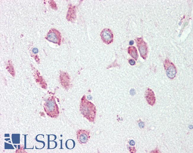 Anti-HEXA antibody IHC staining of human brain, cortex. Immunohistochemistry of formalin-fixed, paraffin-embedded tissue after heat-induced antigen retrieval.
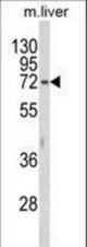 Western blot of HEXA Antibody in mouse liver tissue lysates (35 ug/lane). HEXA (arrow) was detected using the purified antibody.
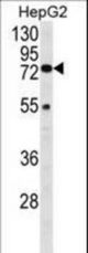 Western blot of HEXA Antibody in HepG2 cell line lysates (35 ug/lane). HEXA (arrow) was detected using the purified antibody.
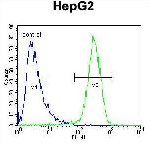 HEXA Antibody flow cytometry of HepG2 cells (right histogram) compared to a negative control cell (left histogram). FITC-conjugated goat-anti-rabbit secondary antibodies were used for the analysis.