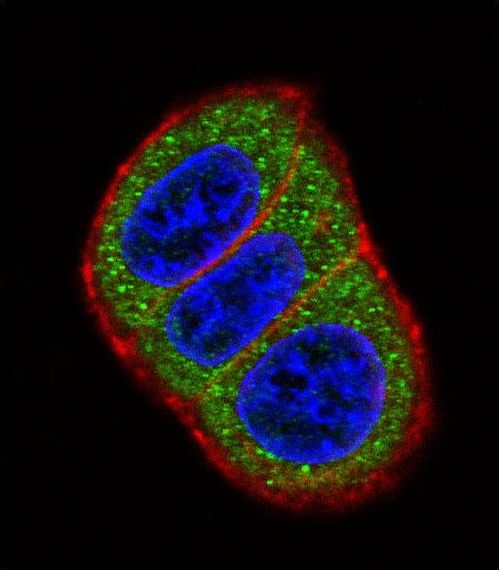
Confocal immunofluorescent of HES1 Antibody (N-term T24) with HeLa cell followed by Alexa Fluor 488-conjugated goat anti-rabbit lgG (green). Actin filaments have been labeled with Alexa Fluor 555 phalloidin (red). DAPI was used to stain the cell nuclear (blue).

Western blot of anti-HES1 Antibody (N-term T24) (RB13980) in HL60 cell line lysates (35 ug/lane). HES1 (arrow) was detected using the purified antibody.

HES1 Antibody (N-term T24) flow cytometry of U251 cells (right histogram) compared to a negative control cell (left histogram). FITC-conjugated goat-anti-rabbit secondary antibodies were used for the analysis.