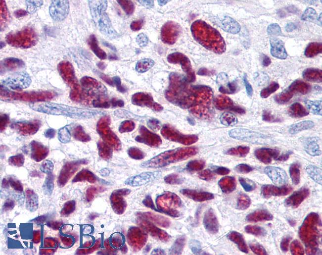
Anti-GLI3 antibody IHC of human brain-glioblastoma. Immunohistochemistry of formalin-fixed, paraffin-embedded tissue after heat-induced antigen retrieval. Antibody LS-B73 concentration 1.25 ug/ml.

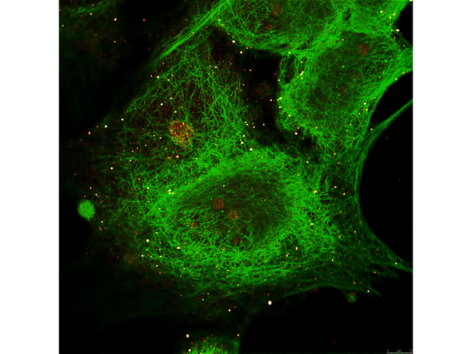
Anti-Gli-3 Antibody - Immunofluorescence. Anti-Gli-3 Antibody Immunofluorescence image showing MCF-7 cell staining of Anti-alpha-Tubulin (MOUSE) Monoclonal Antibody - in green and staining of Anti-Gli-3 (RABBIT) Antibody - LS-B73 in red.

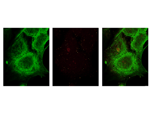
Anti-Gli-3 Antibody - Immunofluorescence. Anti-Gli-3 Antibody Immunofluorescence image one showing MCF-7 cell staining of Anti-alpha-Tubulin (MOUSE) Monoclonal Antibody - in green. Immunofluorescence image two showing MCF-7 cell staining of Anti-Gli-3 (RABBIT) Antibody - LS-B73 in red. Immunofluorescence image three showing MCF-7 cell superimposed staining of Anti-alpha-Tubulin (MOUSE) Monoclonal Antibody - in green and staining of Anti-Gli-3 (RABBIT) Antibody - LS-B73 in red.

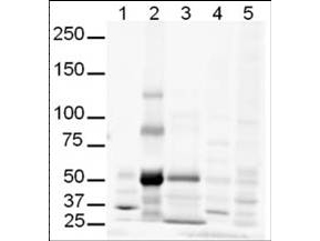
Anti-Gli-3 Antibody - Western Blot. Western blot of anti-Gli-3 antibody shows detection of multiple bands in human lung lysate believed to be Gli-3. Lanes contain 20 ug of whole cell lysates from 1 - human brain, 2 - human lung, 3 - human spleen, 4 - mouse brain and 5 - mouse lung. While no recognizable staining can be seen on mouse tissue, human lung shows what may be truncated Gli-3 (~80kD). This identity of the strong band at ~50 kD is unknown. After blocking, the membrane was probed with the primary antibody diluted to 1:500. For detection use HRP Gt-a-Rabbit IgG (LS-C60865). Detection of Gli-3 by western blot may be enhanced if nuclear extracts are used instead of whole cell lysates as the expression/abundance of Gli-3 is likely to be low. Furthermore, Gli3 expression is likely to be developmentally regulated and induced, making it difficult to detect in whole tissue homogenates.


