ILK1 Antibody
| Name | ILK1 Antibody |
|---|---|
| Supplier | Cell Signaling Technology |
| Catalog | 3862 |
| Prices | $246.00 |
| Sizes | 100 µl (10 western blots) |
| Host | Rabbit |
| Clonality | Polyclonal |
| Applications | WB IHC-P |
| Species Reactivities | Human, Mouse, Rat, Monkey, Bovine |
| Antigen | Polyclonal antibodies are produced by immunizing animals with a synthetic peptide corresponding to the carboxy-terminus of human ILK1 |
| Description | Rabbit Polyclonal |
| Gene | ILK |
| Supplier Page | Shop |
Product images
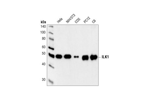 Western blot analysis of extracts from HeLa, NIH/3T3, COS, PC12 and C6 cells, using ILK1 Antibody.
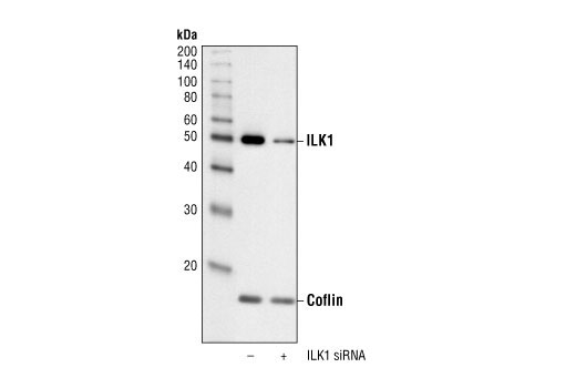 Western blot analysis of extracts from HeLa cells transfected with 50 nM non-targeted siRNA (-) or 100 nM ILK1 siRNA (+), using ILK1 Antibody #3862 and Cofilin Antibody #3312. The ILK1 Antibody confirms silencing of ILK1 expression, and the Cofilin Antibody controls for protein loading and ILK1 siRNA specificity.
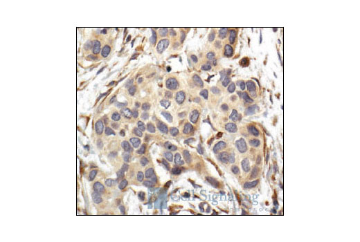 Immunohistochemical analysis of paraffin-embedded human breast carcinoma, showing cytoplasmic localization, using ILK1 Antibody.