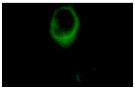
Raf-B (F-7): sc-5284. Immunofluorescence staining of methanol-fixed NIH/3T3 cells showing cytoplasmic localization.
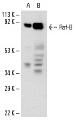
Raf-B (F-7): sc-5284. Western blot analysis of Raf-B expression in HL-60 (A) and NIH/3T3 (B) whole cell lysates.
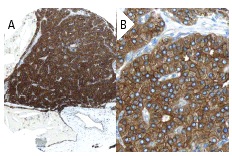
Raf-B (F-7): sc-5284. Immunoperoxidase staining of formalin fixed, paraffin-embedded human parathyroid gland tissue showing cytoplasmic staining of glandular cells (low and high magni?fication). Kindly provided by The Swedish Human Protein Atlas (HPA) program.
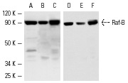
Western blot analysis of Raf B expression in A-431 (A,D), HL-60 (B,E) and NIH/3T3 (C,F) whole cell lysates. Antibodies tested include Raf-B (H-145): sc-9002 (A-C) and Raf-B (F-7): sc-5284 (D-F).