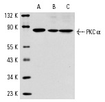
PKC α (C-20)-G: sc-208-G. Western blot analysis of PKC α expression in Jurkat (A) and NIH/3T3 (B) whole cell lysates and rat brain extract (C).
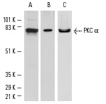
Western blot analysis of PKC α expression in NIH/3T3 (A,C) and Jurkat (B) whole cell lysates. Antibodies tested include PKC α (C-20): sc-208 (A), PKC (MC5): sc-80 (B) and PKC α (H-7): sc-8393 (C).
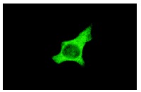
PKC α (C-20): sc-208. Immunofluorescence staining of methanol-fixed HeLa cells.
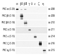
Western blot analysis of Baculovirus expressed PKC isoforms at 10 ng/lane. PKC antibodies used include PKC α (C-20), PKC βI (C-16), PKC βII (C-18), PKC γ (C-19), PKC ε (C-15), PKC ζ (C-20) and PKC η (C-15).
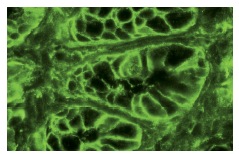
PKC α (C-20): sc-208. Immunofluorescence staining of normal mouse intestine frozen section showing cytoplasmic staining.

Western blot analysis of Akt1 phosphorylation in A-431 cells treated with EGF. Blots were probed with Akt1 (C-20): sc-1618 (A), p-Akt1/2/3 (Ser 473): sc-7985-R preincubated with cognate unphosphorylated peptide (B) and p-Akt1/2/3 (Ser 473): sc-7985-R preincubated with cognate phosphorylated peptide (C).


