Anti-Delta-6 Desaturase / FADS2 Antibody (aa79-108)
| Name | Anti-Delta-6 Desaturase / FADS2 Antibody (aa79-108) |
|---|---|
| Supplier | LifeSpan Bioscience |
| Catalog | LS-C165916 |
| Prices | $295.00 |
| Sizes | 400 µl |
| Host | Rabbit |
| Clonality | Polyclonal |
| Applications | WB ELISA |
| Species Reactivities | Mouse |
| Purity/Format | Protein A purified |
| Blocking Peptide | DZIP1 Antibody Blocking Peptide |
| Description | Rabbit Polyclonal |
| Gene | FADS2 |
| Conjugate | Unconjugated |
| Supplier Page | Shop |
Product images
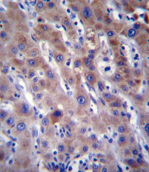 FADS2 Antibody immunohistochemistry of formalin-fixed and paraffin-embedded human liver tissue followed by peroxidase-conjugated secondary antibody and DAB staining.
 Fluorescent image of A549 cell stained with FADS2 Antibody. A549 cells were fixed with 4% PFA (20 min), permeabilized with Triton X-100 (0.1%, 10 min), then incubated with FADS2 primary antibody (1:25, 1 h at 37°C). For secondary antibody, Alexa Fluor 488 conjugated donkey anti-rabbit antibody (green) was used (1:400, 50 min at 37°C). Cytoplasmic actin was counterstained with Alexa Fluor 555 (red) conjugated Phalloidin (7units/ml, 1 h at 37°C). FADS2 immunoreactivity is localized to Cytoplasm and Vesicles significantly.
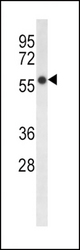 FADS2 Antibody western blot of HepG2 cell line lysates (35 ug/lane). The FADS2 antibody detected the FADS2 protein (arrow).
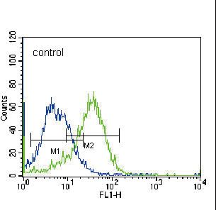 FADS2 Antibody flow cytometry of K562 cells (right histogram) compared to a negative control cell (left histogram). FITC-conjugated donkey-anti-rabbit secondary antibodies were used for the analysis.