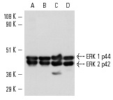
ERK 2 (K-23): sc-153. Western blot analysis of ERK 1 and ERK 2 expression in HeLa (A), A-431 (B), KNRK (C) and NIH/3T3 (D) whole cell lysates.
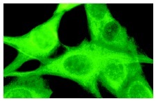
ERK 2 (K-23): sc-153. Immunofluorescence staining of methanol-fixed NIH/3T3 cells showing cytoplasmic localization.
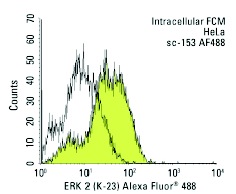
ERK 2 (K-23) Alexa Fluor 488: sc-153 AF488. Intracellular FCM analysis of fixed and permeabilized HeLa cells. Black line histogram represents the isotype control, normal rabbit IgG: sc-45068.
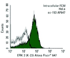
ERK 2 (K-23) Alexa Fluor 647: sc-153 AF647. Intracellular FCM analysis of fixed and permeabilized HeLa cells. Black line histogram represents the isotype control, normal rabbit IgG: sc-24647.
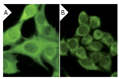
ERK 2 (K-23): sc-153. Immunofluorescence staining of methanol-fixed NIH/3T3 cells showing cytoplasmic localization using indirect FITC (A) staining and HeLa cells using direct Alexa Fluor 488 (B) staining.
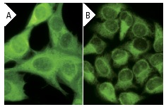
ERK 2 (K-23): sc-153. Immunofluorescence staining of methanol-fixed NIH/3T3 cells showing cytoplasmic localization using indirect FITC (A) staining and HeLa cells using direct Alexa Fluor 488 (B) staining.
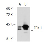
ERK 2 (K-23): sc-153. Western blot analysis of ERK 1 expression in non-transfected: sc-117752 (A) and mouse ERK 1 transfected: sc-126806 (B) 293T whole cell lysates.
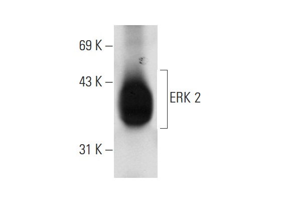
ERK 2 (K-23): sc-153. Western blot analysis of ERK 2 expression in rat hippocampus tissue extract.
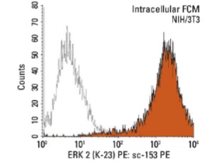
ERK 2 (K-23) PE: sc-153 PE. Intracellular FCM analysis of fixed and permeabilized NIH/3T3 cells. Black line histogram represents the isotype control, normal rabbit IgG: sc-3871.
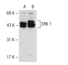
ERK 2 (K-23): sc-153. Western blot analysis of ERK 1 expression in non-transfected: sc-117752 (A) and human ERK 1 transfected: sc-171765 (B) 293T whole cell lysates.
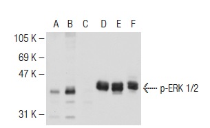
Western blot analysis of ERK 1/2 phosphorylation in untreated (A,D), UV irradiated (B,E) and UV irradiated and lambda protein phosphatase treated (C,F) HeLa whole cell lysates. Antibodies tested include p-ERK 1/2 (Thr 177 )-R: sc-16981-R (A,B,C) and ERK 2 (K-23): sc-153 (D,E,F).
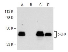
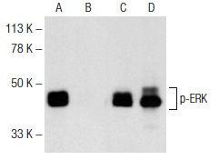
Western blot analysis of ERK phosphorylation in untreated (A,C) and Sorafenib (sc-220125) treated (B,D) SK-MEL-28 whole cell lysates. Antibodies tested include p-ERK (E-4): sc-7383 (A,B) and ERK 2 (K-23): sc-153 (C,D). Note inhibition of ERK phosphorylation by Sorafenib in lane B.
Western blot analysis of ERK phosphorylation in untreated (A,C) and Sorafenib (sc-220125) treated (B,D) SW480 whole cell lysates. Antibodies tested include p-ERK (E-4): sc-7383 (A,B) and ERK 2 (K-23): sc-153 (C,D). Note inhibition of ERK phosphorylation by Sorafenib in lane B.