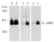
Western blot analysis of inactive (A,C,E) and active (B,D,F) rat recombinant ERK. Antibodies tested include ERK 2 (D-2): sc-1647 (A,B), p-ERK (Tyr 204): sc-7976 (C,D) and p-ERK (Tyr 204)-R: sc-7976-R (E,F).
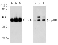
Western blot analysis of unactive rat recombinant ERK 2 (A,D), rat recombinant ERK 2 phosphorylated by MEK-1 (phosphorylated by PDK1) (B,E) and rat recombinant ERK 2 phosphorylated by MEK-1 (phos-phorylated by MEK kinase-1) (C,F). Antibodies tested include ERK 2 (D2): sc-1647 (A-C) and p-ERK (Tyr 204): sc-7976 (D-F).
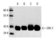
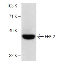
ERK 2 (D-2): sc-1647. Western blot analysis of ERK 2 expression in A-431 (A), HeLa (B), KNRK (C) and NIH/3T3 (D) whole cell lysates.
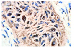
ERK 2 (D-2): sc-1647. Immunoperoxidase staining of formalin-fixed, paraffin-embedded human lung carcinoma tissue showing nuclear and cytoplasmic localization.
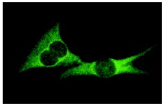
ERK 2 (D-2): sc-1647. Immunofluorescence staining of methanol-fixed NIH/3T3 cells showing cytoplasmic localization.
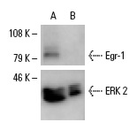
Egr-1 siRNA (h): sc-29303. Western blot analysis of Egr-1 expression in non-transfected control (A) and Egr-1 siRNA transfected (B) EGF-treated A-431 cells. Blot probed with Egr-1 (C-19): sc-189. ERK 2 (D-2): sc-1647 used as specificity and loading control.
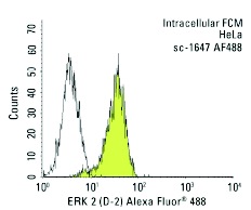
ERK 2 (D-2) Alexa Fluor 488: sc-1647 AF488. Intracellular FCM analysis of fixed and permeabilized HeLa cells. Black line histogram represents the isotype control, normal mouse IgG2b: sc-3892.
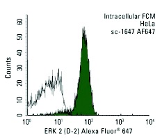
ERK 2 (D-2) Alexa Fluor 647: sc-1647 AF6478. Intracellular FCM analysis of fixed and permeabilized HeLa cells. Black line histogram represents the isotype control, normal mouse IgG2b: sc-24638.
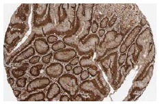
ERK 2 (D-2): sc-1647. Immunoperoxidase staining of formalin fixed, paraffin-embedded human stomach tissue showing cytoplasmic and nuclear staining of glandular cells. Kindly provided by The Swedish Human Protein Atlas (HPA) program.
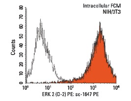
ERK 2 (D-2) PE: sc-1647 PE. Intracellular FCM analysis of fixed and permeabilized NIH/3T3 cells. Black line histogram represents the isotype control, normal mouse IgG
2b: sc-2868.
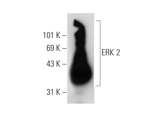
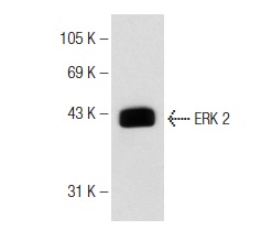
ERK 2 (D-2): sc-1647. Western blot analysis of ERK 2 expression in rat hippocampus tissue extract.
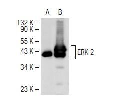
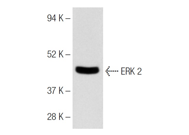
ERK 2 (D-2): sc-1647. Western blot analysis of ERK 2 expression in non-transfected: sc-117752 (A) and human ERK 2 transfected: sc-177196 (B) 293T whole cell lysates.
donkey anti-mouse IgG-HRP: sc-2314. Western blot analysis of ERK 2 expression in Jurkat whole cell lysate. Antibody tested (IgG
2b): ERK 2 (D-2): sc-1647.
donkey anti-mouse IgG-HRP: sc-2096. Western blot analysis of ERK 2 expression in Jurkat whole cell lysate. Antibody tested (IgG
3): ERK 2 (D-2): sc-1647.
goat anti-mouse IgG2b-HRP: sc-2062. Western blot analysis of ERK 2 expression in Jurkat whole cell lysate. Antibody tested: ERK 2 (D-2): sc-1647.
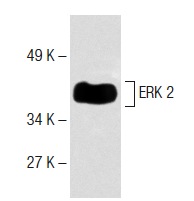
goat anti-mouse IgG
2b-HRP: sc-2971. Western blot analysis of ERK 2 expression in NIH/3T3 whole cell lysate. Antibody tested: ERK 2 (D-2): sc-1647.