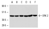
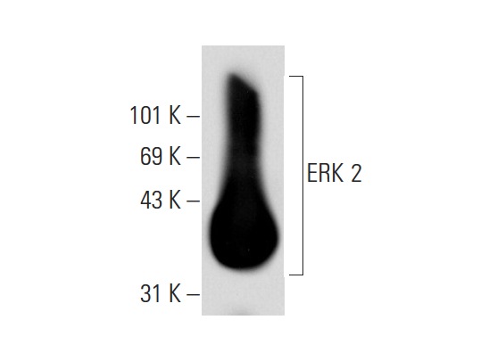
ERK 2 (C-14): sc-154. Western blot analysis of ERK 2 expression in A-431 (A), HeLa (B), KNRK (C), NIH/3T3 (D), NRK (E) and DU 145 (F) whole cell lysates.
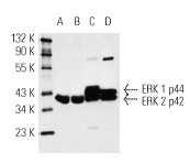
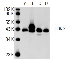
ERK 2 (C-14)-G: sc-154-G. Western blot analysis of ERK 1 and ERK 2 expression in A-431 (A), HeLa (B), KNRK (C) and NIH/3T3 (D) whole cell lysates.
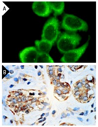
ERK 2 (C-14): sc-154. Immunofluorescence staining of methanol-fixed HeLa cells (A) and immunoperoxidase staining of formalin-fixed, paraffin-embedded human breast carcinoma tissue (B) showing cytoplasmic localization.
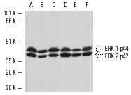
Western blot analysis of ERK in 3611-RF (A,D), NIH/3T3 (B,E) and KNRK (C,F) whole cell lysates. Antibodies tested include ERK 1 (K-23)-G: sc-94-G (A-C) and ERK 2 (C-14)-G: sc-154-G (D-F).
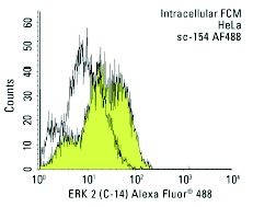
ERK 2 (C-14) Alexa Fluor 488: sc-154 AF488. Intracellular FCM analysis of fixed and permeabilized HeLa cells. Black line histogram represents the isotype control, normal rabbit IgG: sc-45068.
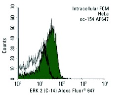
ERK 2 (C-14) Alexa Fluor 647: sc-154 AF647. Intracellular FCM analysis of fixed and permeabilized HeLa cells. Black line histogram represents the isotype control, normal rabbit IgG: sc-24647.
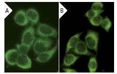
ERK 2 (C-14): sc-154. Immunofluorescence staining of methanol-fixed HeLa cells showing cytoplasmic localization using indirect FITC (A) staining and direct Alexa Fluor 488 (B) staining.
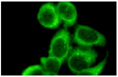
ERK 2 (C-14): sc-154. Immunofluorescence staining of methanol-fixed HeLa cells showing cytoplasmic localization.
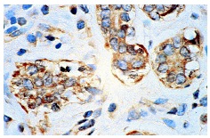
ERK 2 (C-14): sc-154. Immunoperoxidase staining of formalin-fixed, paraffin-embedded human breast carcinoma tissue showing cytoplasmic localization.
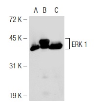
ERK 2 (C-14): sc-154. Western blot analysis of ERK 1 expression in non-transfected 293T: sc-117752 (A), mouse ERK 1 transfected 293T: sc-126806 (B) and A-431 (C) whole cell lysates.
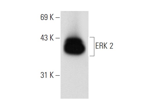
ERK 2 (C-14): sc-154. Western blot analysis of ERK 2 expression in rat hippocampus tissue extract.
ERK 2 (C-14)-G: sc-154-G. Western blot analysis of ERK 2 expression in rat hippocampus tissue extract.
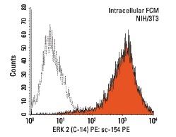
ERK 2 (C-14) PE: sc-154 PE. Intracellular FCM analysis of fixed and permeabilized NIH/3T3 cells. Black line histogram represents the isotype control, normal rabbit IgG: sc-3871.
ERK 2 (C-14)-G: sc-154-G. Western blot analysis of ERK 2 expression in non-transfected 293T: sc-117752 (A), human ERK 2 transfected 293T: sc-177196 (B), A-431 (C) and HeLa (D) whole cell lysates.
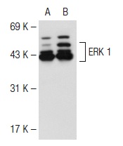
ERK 1 (C-14): sc-154. Western blot analysis of ERK 1 expression in non-transfected: sc-117752 (A) and human ERK 1 transfected: sc-171765 (B) 293T whole cell lysates.