
Anti-DAPK2 / DRP1 antibody IHC staining of human heart. Immunohistochemistry of formalin-fixed, paraffin-embedded tissue after heat-induced antigen retrieval. Antibody LS-B9958 concentration 10 ug/ml.
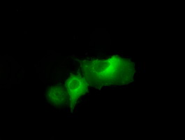
Anti-DAPK2 mouse monoclonal antibody immunofluorescent staining of COS7 cells transiently transfected by pCMV6-ENTRY DAPK2.

HEK293T cells were transfected with the pCMV6-ENTRY control (Left lane) or pCMV6-ENTRY DAPK2 (Right lane) cDNA for 48 hrs and lysed. Equivalent amounts of cell lysates (5 ug per lane) were separated by SDS-PAGE and immunoblotted with anti-DAPK2.

Immunoprecipitation(IP) of DAPK2 by using monoclonal anti-DAPK2 antibodies (Negative control: IP without adding anti-DAPK2 antibody.). For each experiment, 500ul of DDK tagged DAPK2 overexpression lysates (at 1:5 dilution with HEK293T lysate), 2 ug of anti-DAPK2 antibody and 20ul (0.1 mg) of goat anti-mouse conjugated magnetic beads were mixed and incubated overnight. After extensive wash to remove any non-specific binding, the immuno-precipitated products were analyzed with rabbit anti-DDK polyclonal antibody.