Anti-Neurogenin-3 (rabbit) Antibody
| Name | Anti-Neurogenin-3 (rabbit) Antibody |
|---|---|
| Supplier | Sigma-Aldrich |
| Catalog | AB10535 |
| Prices | $319.00 |
| Sizes | ab10535 |
| Clonality | Polyclonal |
| Isotype | IgG |
| Applications | WB |
| Species Reactivities | Mouse |
| Purity/Format | Unpurified |
| Description | Polyclonal |
| Gene | Neurog3 |
| Supplier Page | Shop |
Product images
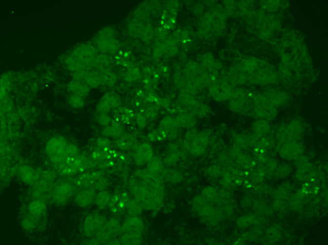 Immunofluorescent Analysis: Staining pattern and morphology in e15.5 mouse pancreas. Fixed frozen tissue, no antigen retrieval. Antibody diluted 1:2000; secondary detection method was fluorescence (alexa488 conjugated donkey anti-rabbit).
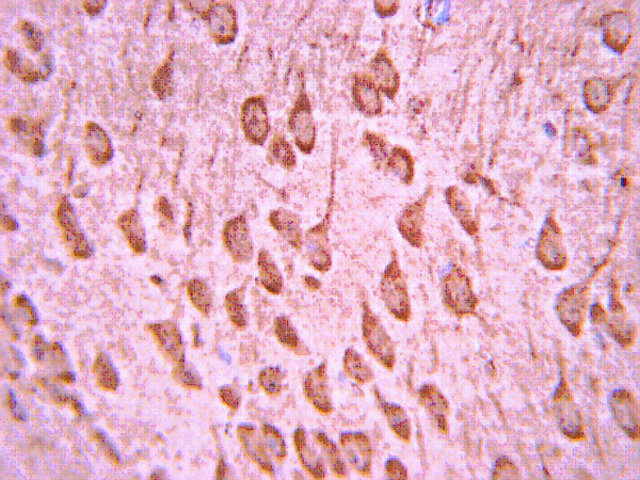 Immunohistochemistry Analysis:
 Representative lot data.
Paraffin-embedded mouse embryo E12.5+ tissue was prepared using heat-induced epitope retrieval in citrate buffer, pH 6.0. Immunostaining was performed using a 1:500 dilution of Cat. No. AB10535, Anti-Neurogenin-3 (rabbit polyclonal). Reactivity was detected using the IHC-Select Detection Kit (Cat. No. DAB050). Immunoreactivity is expressed in embryonic brain in synaptic processes. Staining pattern apears to be cytoplasmic.
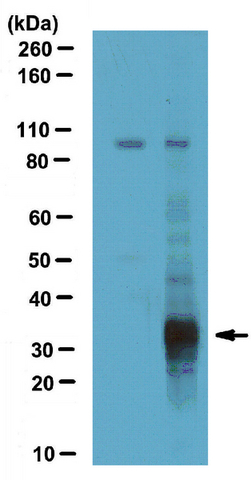 Western Blotting Analysis:
Representative lot data.
mNgn3 transfected (lane 2) and wildtype (lane 1) rabbit reticulocyte lysates were resolved by electrophoresis, transferred to PVDF membranes and probed with a 1:1,000 dilution of Anti-Neurogenin-3 (rabbit polyclonal) antibody.
Proteins were visualized using a Donkey Anti-Rabbit IgG conjugated to HRP and chemiluminescence detection system.
Arrow indicates Neurogenin-3 (~32 kDa).