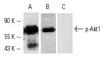
Western blot analysis of Akt1 phosphorylation in A-431 cells treated with EGF. Blots were probed with Akt1 (C-20): sc-1618 (A), p-Akt1/2/3 (Ser 473): sc-7985-R preincubated with cognate unphosphorylated peptide (B) and p-Akt1/2/3 (Ser 473): sc-7985-R preincubated with cognate phosphorylated peptide (C).
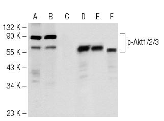
Western blot analysis of Akt1/2/3 phosphorylation in untreated (A, D), calyculin treated (B, E) and calyculin and lambda protein phosphatase treated (C, F) Jurkat whole cell lysates. Antibodies tested include p-Akt1/2/3 (Ser 473)-R: sc-7985-R (A, B, C) and Akt1 (C-20): sc-1618 (D, E, F).
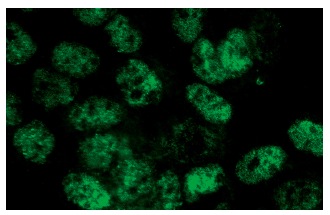
p-Akt1/2/3 (Ser 473)-R: sc-7985-R. Immunofluorescence staining of methanol-fixed A-431 cells showing nuclear localization.
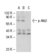
p-Akt1/2/3 (Ser 473)-R: sc-7985-R. Western blot analysis of Akt2 phosphorylation in non-transfected: sc-117752 (A), untreated human Akt2 transfected: sc-116831 (B) and lambda protein phosphatase treated human Akt2 transfected: sc-116831 (C) 293T whole cell lysates.
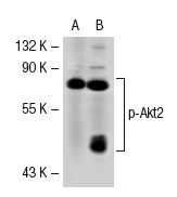
p-Akt1/2/3 (Ser 473)-R: sc-7985-R. Western blot analysis of Akt2 phosphorylation in non-transfected: sc-117752 (A) and human Akt2 transfected: sc-116831 (B) 293T whole cell lysates.
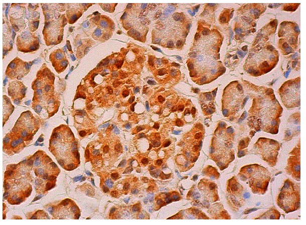
p-Akt1/2/3 (Ser 473)-R: sc-7985-R. Immunoperoxidase staining of formalin fixed, paraffin-embedded human pancreas tissue showing nuclear and cytoplasmic staining of exocrine glandular cells and Islets of Langerhans.