Anti-CTSE / Cathepsin E Antibody (Internal)
| Name | Anti-CTSE / Cathepsin E Antibody (Internal) |
|---|---|
| Supplier | LifeSpan Bioscience |
| Catalog | LS-C99298 |
| Host | Rabbit |
| Clonality | Polyclonal |
| Applications | IHC-P WB FC |
| Species Reactivities | Human, Mouse |
| Antigen | CTSE / Cathepsin E antibody was raised against kLH conjugated synthetic peptide selected from the Center region of human CTSE |
| Blocking Peptide | CTSD / Cathepsin D Antibody Blocking Peptide |
| Description | Rabbit Polyclonal |
| Gene | CTSE |
| Conjugate | Unconjugated |
| Supplier Page | Shop |
Product images
 CTSE Antibody IHC of formalin-fixed and paraffin-embedded human lung carcinoma followed by peroxidase-conjugated secondary antibody and DAB staining.
 Western blot of CTSE antibody in mouse stomach tissue lysates (35 ug/lane). CTSE(arrow) was detected using the purified antibody.
 Western blot of CTSE antibody in K562 cell line lysates (35 ug/lane). CTSE(arrow) was detected using the purified antibody.
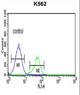 CTSE Antibody flow cytometry of K562 cells (right histogram) compared to a negative control cell (left histogram). FITC-conjugated goat-anti-rabbit secondary antibodies were used for the analysis.