Fluorescent confocal image of SK-BR-3 cell stained with CTBP1 Antibody. SK-BR-3 cells were fixed with 4% PFA (20 min), permeabilized with Triton X-100 (0.1%, 10 min), then incubated with CTBP1 primary antibody (1:25, 1 h at 37°C). For secondary antibody, Alexa Fluor 488 conjugated donkey anti-rabbit antibody (green) was used (1:400, 50 min at 37°C). Cytoplasmic actin was counterstained with Alexa Fluor 555 (red) conjugated Phalloidin (7units/ml, 1 h at 37°C). Nuclei were counterstained with DAPI (blue) (10 ug/ml, 10 min). CTBP1 immunoreactivity is localized to nucleus significantly.

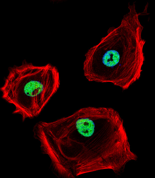
Fluorescent confocal image of HeLa cell stained with CTBP1 Antibody. HeLa cells were fixed with 4% PFA (20 min), permeabilized with Triton X-100 (0.1%, 10 min), then incubated with CTBP1 primary antibody (1:25, 1 h at 37°C). For secondary antibody, Alexa Fluor 488 conjugated donkey anti-rabbit antibody (green) was used (1:400, 50 min at 37°C). Cytoplasmic actin was counterstained with Alexa Fluor 555 (red) conjugated Phalloidin (7units/ml, 1 h at 37°C). Nuclei were counterstained with DAPI (blue) (10 ug/ml, 10 min). CTBP1 immunoreactivity is localized to nucleus significantly.
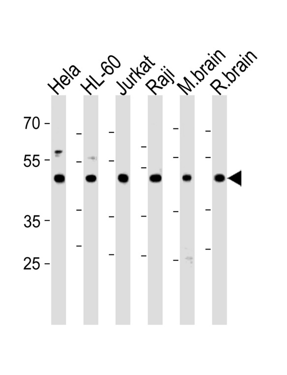
CTBP1 Antibody western blot of HeLa,HL-60,Jurkat,Raji cell line and mouse brain,rat brain tissue lysates (35 ug/lane). The CTBP1 antibody detected the CTBP1 protein (arrow).