Western blot analysis of Aurora A was performed by loading 30 ug of Jurkat (lane1), K562 (lane2) and MCF7 (lane3) cell lysate using Novex® NuPAGE® 4-12 % Bis-Tris gel (NP0321BOX), XCell SureLock™ Electrophoresis System (EI0002), Novex® Sharp Pre-Stained Protein Standard (LC5800), and iBlot® Dry Blotting System (IB21001). Proteins were transferred to a nitrocellulose membrane and blocked with 5 % skim milk for 1 hour at room temperature. Aurora A was detected at ~ 45 kDa using Aurora A Mouse Monoclonal Antibody (458900) at 1-3 ug/ml in 5 % skim milk at 4°C overnight on a rocking platform. Goat Anti-Mouse IgG - HRP Secondary Antibody (626520) 1:4000 dilution was used and chemiluminescent detection was performed using Novex® ECL Chemiluminescent Substrate Reagent Kit (WP20005).

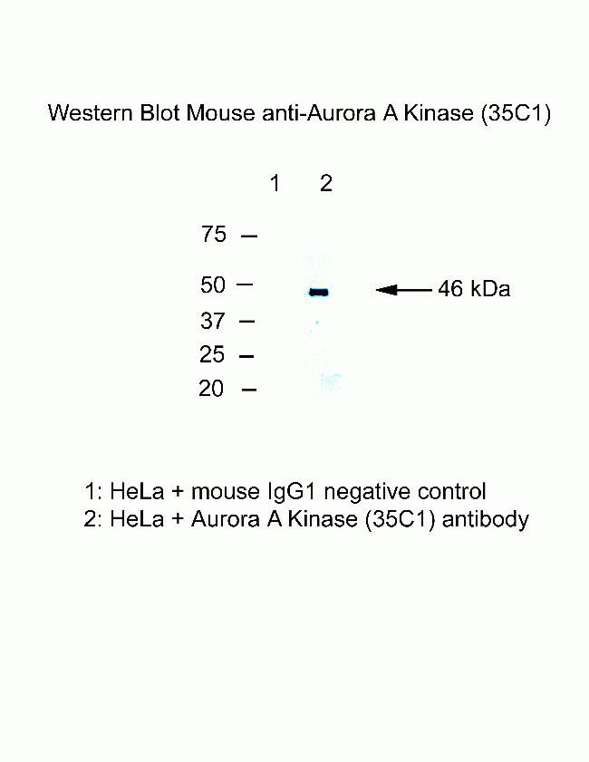
Western blot of Aurora-A kinase

Immunofluorescence analysis of Aurora A Antibody (35C1)was done on 70% confluent log phase HeLa cells. The cells were fixed with 4% paraformaldehyde for 15 minutes, permeabilized with 0.25% Triton™ X-100 for 10 minutes, and blocked with 5% BSA for 1 hour at room temperature. The cells were labeled with Aurora A Antibody (35C1)(458900) at 1µg/mL in 1% BSA and incubated for 3 hours at room temperature and then labeled with Alexa Flour 488 Rabbit Anti-Mouse IgG Secondary Antibody (A11059) at a dilution of 1:400 for 45 minutes at room temperature (Panel a: green). Nuclei (Panel b: blue) were stained with SlowFade® Gold Antifade Mountant with DAPI (S36938). F-actin (Panel c: red) was stained with Alexa Fluor 594 Phalloidin (A12381). Panel d is a merged image showing cytoplasmic and Nuclear localization. Panel e is a no primary antibody control. The images were captured at 40X magnification.


