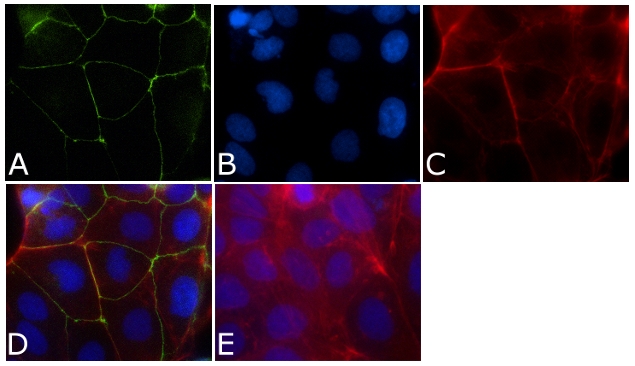
Immunofluorescence analysis of ZO-1 / TJP1 Antibody, FITC conjugate (ZO1-1A12) was done on 90% confluent log phase CaCo2 cells. The cells were fixed with 4% paraformaldehyde for 15 minutes, permeabilized with 0.25% Triton™ X-100 for 10 minutes, and blocked with 5% BSA for 1 hour at room temperature. The cells were labeled with ZO-1 / TJP1 Antibody, FITC conjugate (ZO1-1A12)(339111) at 1µg/mL in 1% BSA and incubated for 3 hours at room temperature (Panel a: green). Nuclei (Panel b: blue) were stained with SlowFade® Gold Antifade Mountant with DAPI (S36938). F-actin (Panel c: red) was stained with Alexa Fluor 594 Phalloidin (A12381). Panel d is a merged image showing cell junctional localization. Panel e is a no primary antibody control. The images were captured at 40X magnification.