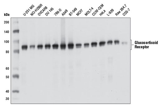
Western blot analysis of extracts from various cell lines using Glucocorticoid Receptor (D6H2L) XP ® Rabbit mAb.

Western blot analysis of extracts from 293T cells, either mock transfected (-) or transfected with constructs expressing Myc/DDK-tagged full-length human glucocorticoid receptor-α (hGRα-Myc/DDK; +) or Myc/DDK-tagged full-length human mineralocorticoid receptor (hMR-Myc/DDK; +), using Glucocorticoid Receptor (D6H2L) XP ® Rabbit mAb (upper) or DYKDDDDK Tag Antibody #2368 (lower).

Immunoprecipitation of glucocorticoid receptor from HeLa cell extracts using Rabbit (DA1E) mAb IgG XP ® Isotype Control #3900 (lane 2) or Glucocorticoid Receptor (D6H2L) XP ® Rabbit mAb (lane 3). Lane 1 is 10% input. Western blot analysis was performed using Glucocorticoid Receptor (D6H2L) XP ® Rabbit mAb.

Immunohistochemical analysis of paraffin-embedded human colon carcinoma using Glucocorticoid Receptor (D6H2L) XP ® Rabbit mAb.

Immunohistochemical analysis of paraffin-embedded human lung carcinoma using Glucocorticoid Receptor (D6H2L) XP ® Rabbit mAb.

Immunohistochemical analysis of paraffin-embedded mouse stomach using Glucocorticoid Receptor (D6H2L) XP ® Rabbit mAb.

Immunohistochemical analysis of paraffin-embedded HeLa cell pellets, untreated (left) or dexamethasone-treated (right), using Glucocorticoid Receptor (D6H2L) XP ® Rabbit mAb.

Immunohistochemical analysis of paraffin-embedded human prostate carcinoma using Glucocorticoid Receptor (D6H2L) XP ® Rabbit mAb.

Confocal immunofluorescent analysis of HeLa cells, grown in phenol red-free media containing 5% charcoal-stripped FBS for 2 d and either untreated (left) or treated with dexamethasone (100 nM, 2 hr; right), using Glucocorticoid Receptor (D6H2L) XP ® Rabbit mAb (green). Actin filaments were labeled with DY-554 phalloidin (red).

Human whole blood was fixed, lysed, and permeabilized as per the Cell Signaling Technology Flow Cytometry (Alternate) Protocol, and stained with CD3-PE, CD19-APC, and Glucocorticoid Receptor (D6H2L) XP ® Rabbit mAb. B cell (green) and T cell (blue) population gates (left) were applied to a histogram depicting the mean fluorescence intensity of glucocorticoid receptor, compared to a nonspecific negative control antibody (red; right). Anti-rabbit IgG (H+L), F(ab') 2 Fragment (Alexa Fluor ® 488 Conjugate) #4412 was used as a secondary antibody.

Chromatin immunoprecipitations were performed with cross-linked chromatin from 4 x 10 6 A549 cells cultured in media with 5% charcoal-stripped FBS for 3 d and then treated with 100 nM dexamethasone for 1 hr and 10 µl of Glucocorticoid Receptor (D6H2L) XP ® Rabbit mAb, using SimpleChIP ® Enzymatic Chromatin IP Kit (Magnetic Beads) #9003. DNA Libraries were prepared from 5 ng enriched ChIP DNA using NEBNext ® Ultra™ II DNA Library Prep Kit for Illumina ® , and sequenced on the Illumina NextSeq. The figure shows binding across SLC19A2, a known target gene of GR (see additional figure containing ChIP-qPCR data). For additional ChIP-seq tracks, please download the product data sheet.

A549 cells were cultured in media with 5% charcoal-stripped FBS for 3 d and then either untreated (left panel) or dexamethasone-treated (100 nM, 1 hr; right panel). Chromatin immunoprecipitations were performed with cross-linked chromatin from 4 x 10 6 cells and 10 µl of Glucocorticoid Receptor (D6H2L) XP ® Rabbit mAb or 2 µl of Normal Rabbit IgG #2729 using SimpleChIP ® Enzymatic Chromatin IP Kit (Magnetic Beads) #9003. The enriched DNA was quantified by real-time PCR using SimpleChIP ® Human SLC19A2 Promoter Primers #7681, human MT2A promoter primers, and SimpleChIP ® Human α Satellite Repeat Primers #4486. The amount of immunoprecipitated DNA in each sample is represented as signal relative to the total amount of input chromatin, which is equivalent to one.