
CB2 antibody immunohistochemistry of formalin-fixed and paraffin-embedded human skin carcinoma followed by peroxidase-conjugated secondary antibody and DAB staining.

Immunohistochemical of paraffin-embedded H. brain section using CB2 Antibody. Antibody was diluted at 1:25 dilution. A peroxidase-conjugated goat anti-rabbit IgG at 1:400 dilution was used as the secondary antibody, followed by DAB staining.

Immunohistochemical of paraffin-embedded R. brain section using CB2 Antibody. Antibody was diluted at 1:25 dilution. A peroxidase-conjugated goat anti-rabbit IgG at 1:400 dilution was used as the secondary antibody, followed by DAB staining.

CB2 Antibody western blot of A431 cell line lysates (35 ug/lane). The CB2 antibody detected the CB2 protein (arrow).

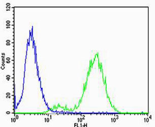
CB2 Antibody flow cytometry of Jurkat cells (right histogram) compared to a negative control cell (left histogram). FITC-conjugated goat-anti-rabbit secondary antibodies were used for the analysis.
Flow cytometric of Jurkat cells using Park7 (DJ-1) Antibody(green) compared to an isotype control of rabbit IgG(blue). Antibody was diluted at 1:25 dilution. An Alexa Fluor 488 goat anti-rabbit lgG at 1:400 dilution was used as the secondary antibody.