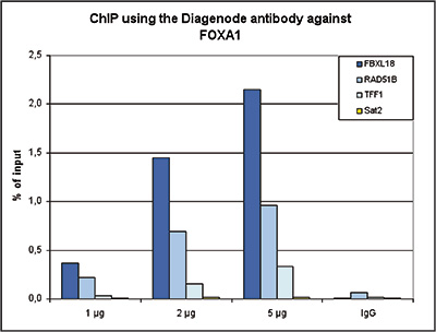
 Figure 1. ChIP results obtained with the Diagenode antibody directed against FOXA1
 ChIP assays were performed using HepG2 cells, the Diagenode antibody against FOXA1 Cat. No. C15410231) and optimized primer sets for qPCR. ChIP was performed with the “iDeal ChIP-seq” kit (Cat. No. C01010055), using sheared chromatin from 4 million cells. A titration of the antibody consisting of 1, 2 and 5 μg per ChIP experiment was analysed. IgG (1 μg/IP) was used as negative IP control. QPCR was performed with primers for the FBXL18, RAD51B and TFF1 genes, used as positive controls, and for the Sat2 satellite repeat, used as a negative control. Figure 1 shows the recovery, expressed as a % of input (the relative amount of immunoprecipitated DNA compared to input DNA after qPCR analysis).
 Figure 2. ChIP-seq results obtained with the Diagenode antibody directed against FOXA1
 ChIP was performed on sheared chromatin from 4 million HepG2 cells using 2 μg of the Diagenode antibody against FOXA1 (Cat. No. C15410231) as described above. The IP’d DNA was subsequently analysed on an Illumina HiSeq. Library preparation, cluster generation and sequencing were performed according to the manufacturer’s instructions. The 50 bp tags were aligned to the human genome using the BWA algorithm. Figure 2 shows the enrichment along the complete sequence and an 800 kb region of human chromosome 1 (fig 2A and B), and in two genomic regions surrounding the FBXL18 and RAD51B positive control genes (fig 2C and D). The position of the amplicon used for qPCR ChIP validation is indicated by an arrow.
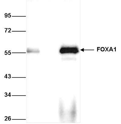
 Figure 3. Western blot analysis using the Diagenode antibody directed against FOXA1
 Nuclear extracts from HepG2 cells (30 μg) were analysed by Western blot using the Diagenode antibody against FOXA1 (Cat. No. C15410231) diluted 1:1,000. The position of the protein of interest is indicated on the right; the marker (in kDa) is shown on the left.
 Figure 4. Immunoprecipitation using the Diagenode antibody directed against FOXA1
 Immunoprecipitation was performed on whole cell extracts from HepG2 cells using 2.5 μg of the Diagenode antibody against FOXA1 (Cat. No. C15410231). An equal amount of rabbit IgG was used as a negative control. The immunoprecipitated FOXA1 protein was detected by western blot with the FOXA1 antibody diluted 1:1,000. The IP with the FOXA1 antibody and with the IgG negative control are shown in lane 3 and lane 2, respectively. Lane 1 shows the input (40 μg of HepG2 whole cell extract).
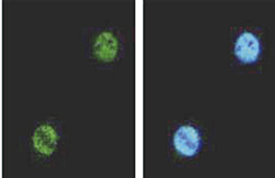
 Figure 5. Immunofluorescence with the Diagenode antibody directed against FOXA1
 HeLa cells were fixed with formaldehyde and stained with the Diagenode antibody against FOXA1 (Cat. C15410231) diluted 1:1,000 (left). The right picture shows costaining with Hoechst 33342 nucleic acid stain.
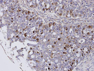
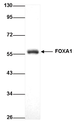 Figure 6. Immunohistochemistry using the Diagenode antibody directed against FOXA1
 Formalin fixed paraffin embedded cervical carcinoma (fig 6A) or breast carcinoma (fig 6B) tissue was stained with the Diagenode antibody against FOXA1 (Cat. No. C15410231) diluted 1:500 followed by a peroxidase labelled goat anti- rabbit secondary antibody.


