Anti-CEP70 Antibody (aa316-346)
| Name | Anti-CEP70 Antibody (aa316-346) |
|---|---|
| Supplier | LifeSpan Bioscience |
| Catalog | LS-C168681 |
| Prices | $295.00 |
| Sizes | 400 µl |
| Host | Rabbit |
| Clonality | Polyclonal |
| Applications | IHC-P WB FC |
| Species Reactivities | Human, Hamster |
| Antigen | CEP70 antibody was raised against kLH-conjugated synthetic peptide from internal region of human CEP70. |
| Purity/Format | Immunoaffinity purified |
| Blocking Peptide | CEP104 Antibody Blocking Peptide |
| Description | Rabbit Polyclonal |
| Gene | CEP70 |
| Supplier Page | Shop |
Product images
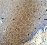 CEP70 antibody immunohistochemistry of formalin-fixed and paraffin-embedded human skin carcinoma followed by peroxidase-conjugated secondary antibody and DAB staining.
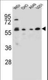 CEP70 Antibody western blot of WiDr,CHO,A549,U251 cell line lysates (35 ug/lane). The CEP70 antibody detected the CEP70 protein (arrow).
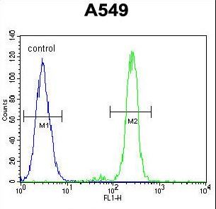 CEP70 Antibody flow cytometry of A549 cells (right histogram) compared to a negative control cell (left histogram). FITC-conjugated goat-anti-rabbit secondary antibodies were used for the analysis.