Anti-CDH20 / Cadherin 20 Antibody
| Name | Anti-CDH20 / Cadherin 20 Antibody |
|---|---|
| Supplier | LifeSpan Bioscience |
| Catalog | LS-C168608 |
| Prices | $295.00 |
| Sizes | 400 µl |
| Host | Rabbit |
| Clonality | Polyclonal |
| Applications | IHC-P WB FC |
| Species Reactivities | Human |
| Antigen | CDH20 / Cadherin 20 antibody was raised against kLH-conjugated synthetic peptide selected from N-terminal region of human CDH20. |
| Purity/Format | Immunoaffinity purified |
| Blocking Peptide | CDH11 / Cadherin 11 Antibody Blocking Peptide |
| Description | Rabbit Polyclonal |
| Gene | CDH20 |
| Supplier Page | Shop |
Product images
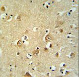 CDH20 Antibody IHC of formalin-fixed and paraffin-embedded brain tissue followed by peroxidase-conjugated secondary antibody and DAB staining.
 Western blot of CDH20 Antibody in HepG2 cell line lysates (35 ug/lane). CDH20 (arrow) was detected using the purified antibody.
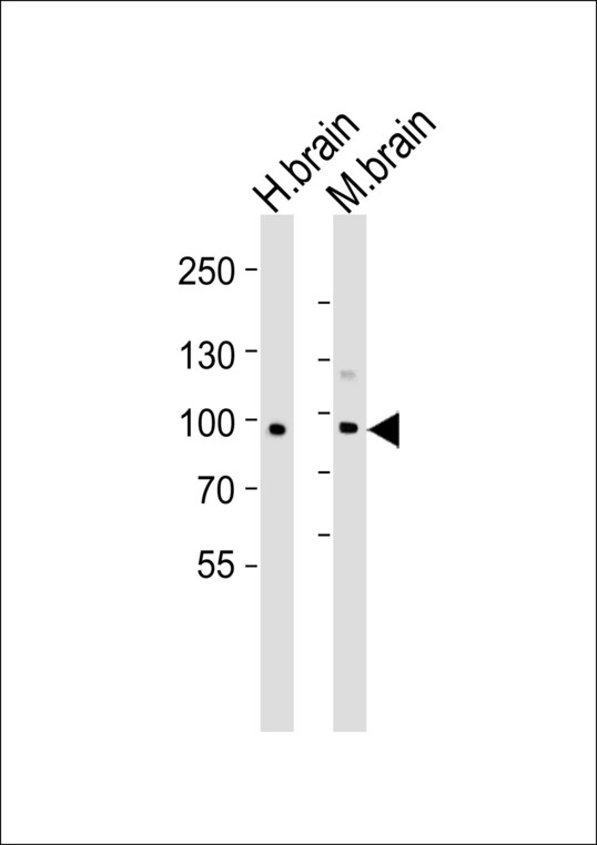 Western blot of lysates from human brain and mouse brain tissue lysate(from left to right), using CDH20 Antibody. Antibody was diluted at 1:1000 at each lane. A goat anti-rabbit IgG H&L (HRP) at 1:5000 dilution was used as the secondary antibody. Lysates at 35ug per lane.
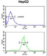 CDH20 Antibody flow cytometry of HepG2 cells (bottom histogram) compared to a negative control cell (top histogram). FITC-conjugated goat-anti-rabbit secondary antibodies were used for the analysis.