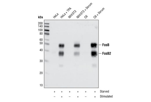
Western blot analysis of extracts from HeLa cells serum-starved overnight and TPA-stimulated for 4 hours, or NIH/3T3 cells and C6 cells serum-starved overnight and serum-stimulated for 4 hours, using FosB (5G4) Rabbit mAb.

Immunohistochemical analysis of paraffin-embedded human breast carcinoma, using FosB (5G4) Rabbit mAb.

Immunohistochemical analysis of paraffin-embedded human breast carcinoma, using FosB (5G4) Rabbit mAb in the presence of control peptide (left) or FosB Blocking Peptide #1042 (right).

Immunohistochemical analysis of paraffin-embedded HeLa cells control (left) or PMA-treated (right), using FosB (5G4) Rabbit mAb.

Confocal immunofluorescent analysis of HeLa cells either serum-starved (left) or TPA-treated (right) and labeled with FosB (5G4) Rabbit mAb (green). Actin filaments have been labeled with Alexa Fluor® 555 phalloidin (red).

Flow cytometric analysis of HeLa cells, untreated (blue) or TPA treated (green), using FosB (5G4) Rabbit mAb compared to a nonspecific negative control antibody (red).

Chromatin immunoprecipitations were performed with cross-linked chromatin from 4 x 10 6 PC-12 cells starved overnight and treated with β-NGF #5221 (50 ng/ml) for 2 hr and 10 μl of FosB (5G4) Rabbit mAb, using SimpleChIP ® Enzymatic Chromatin IP Kit (Magnetic Beads) #9005. DNA Libraries were prepared from 5 ng enriched ChIP DNA using NEBNext ® Ultra™ II DNA Library Prep Kit for Illumina ® , and sequenced on the Illumina NextSeq. The figure shows binding across Ccrn4l, a known target gene of FosB (see additional figure containing ChIP-qPCR data). For additional ChIP-seq tracks, please download the product data sheet.

Chromatin immunoprecipitations were performed with cross-linked chromatin from 4 x 10 6 PC-12 cells starved overnight and treated with β-NGF #5221 (50 ng/ml) for 2 hr and either 10 μl of FosB (5G4) Rabbit mAb or 2 μl of Normal Rabbit IgG #2729 using SimpleChIP ® Enzymatic Chromatin IP Kit (Magnetic Beads) #9003. The enriched DNA was quantified by real-time PCR SimpleChIP ® Rat CCRN4L Promoter Primers #7983, rat DCLK1 promoter primers, and SimpleChIP ® Rat GAPDH Promoter Primers #7964. The amount of immunoprecipitated DNA in each sample is represented as signal relative to the total amount of input chromatin, which is equivalent to one.