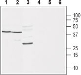
Western blot analysis of rat brain (lanes 1 and 4), mouse brain (lanes 2 and 5) and rat skeletal muscle (lanes 3 and 6): 1-3. Anti-Na V β 1 (extracellular) antibody (#WGA1238), (1:400). 4-6. Anti-Na V β 1 (extracellular) antibody, preincubated with the control peptide antigen.
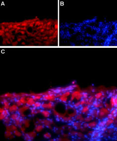
Expression of Na V β1 in rat DRG Immunohistochemical staining of adult rat dorsal root ganglion (DRG) using Anti- Na V β 1 (extracellular) antibody (#WGA1238) followed by goat anti-rabbit-AlexaFluor-594 secondary antibody. A. Na v β1 labeling (red) appears in the cell bodies of the DRG neurons. B. Nuclear staining using DAPI as the counterstain (blue). C. Merged image of A and B.
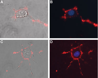
Expression of Na V β 1 in differentiated rat PC12 cells Immunocytochemical staining of live and intact differentiated rat PC12 cells. PC12 differentiation was induced by mNGF 2.5S (>95%) (#N-100). A, C. Merge of Na V β 1 staining using Anti-Na V β 1 (extracellular) antibody (#WGA1238), (1:50), (red) with live cell imaging. B, D. Merge of Na V β 1 staining (1:50), (red) with nuclear staining using DAPI as the counterstain.