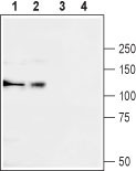
Western blot analysis of rat brain (lanes 1 and 3) and K562 (lanes 2 and 4) lysates: 1, 2. Anti-Ca V α2δ3 (extracellular) antibody (#WGA1247), (1:200). 3, 4. Anti-Ca V α2δ3 (extracellular) antibody preincubated with the control peptide antigen.
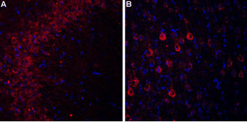
Expression of Cavα2δ3 in rat hippocampus and cortex Immunohistochemical staining of rat hippocampal CA3 region (A) and rat neocortex (B) using Anti-Cavα2δ3 (extracellular) antibody (#WGA1247). In both areas Cavα2δ3 staining (red) appears in pyramidal neurons (arrow). DAPI is used as the counterstain (blue).
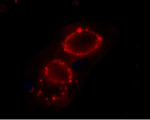
Expression of Ca V α2δ3 in rat PC12 cells Immunocytochemical staining of intact living PC12 cells. Extracellular staining of cells using Anti-Ca v α2δ3 (extracellular) antibody (#WGA1247), (1:25) followed by goat anti-rabbit-AlexaFluor-594 secondary antibody.