Anti-CAV3 / Caveolin 3 Antibody (aa5-31)
| Name | Anti-CAV3 / Caveolin 3 Antibody (aa5-31) |
|---|---|
| Supplier | LifeSpan Bioscience |
| Catalog | LS-C99234 |
| Prices | $295.00 |
| Sizes | 400 µl |
| Host | Rabbit |
| Clonality | Polyclonal |
| Applications | IHC-P WB |
| Species Reactivities | Human, Monkey, Mouse, Rat, Bovine, Horse, Rabbit |
| Purity/Format | Ammonium sulfate precipitation |
| Blocking Peptide | CATSPER2 Antibody Blocking Peptide |
| Description | Rabbit Polyclonal |
| Gene | CAV3 |
| Conjugate | Unconjugated |
| Supplier Page | Shop |
Product images
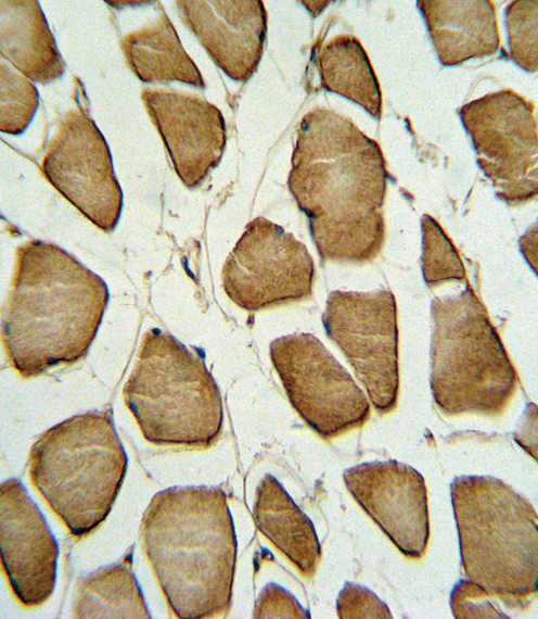 Formalin-fixed and paraffin-embedded human Skeletal muscle reacted with CAV3 Antibody , which was peroxidase-conjugated to the secondary antibody, followed by DAB staining. This data demonstrates the use of this antibody for immunohistochemistry; clinical relevance has not been evaluated.
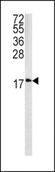 Western blot of CAV3 antibody in 293 cell line lysates (35 ug/lane). CAV3 (arrow) was detected using the purified antibody.
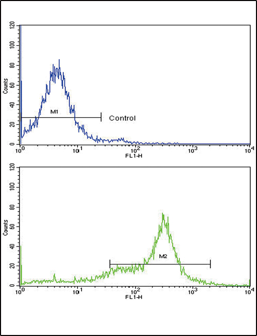 Flow cytometric of 293 cells using CAV3 Antibody (bottom histogram) compared to a negative control cell (top histogram). FITC-conjugated goat-anti-rabbit secondary antibodies were used for the analysis.