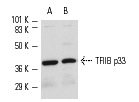
TFIIB (C-18): sc-225. Western blot analysis of TFIIB p33 expression in Jurkat (A) and K-562 (B) whole cell lysates.
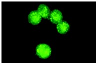
TFIIB (C-18): sc-225. Immunofluorescence staining of methanol-fixed K-562 cells showing nuclear staining.
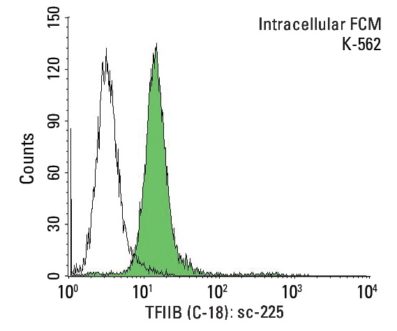
TFIIB (C-18): sc-225. Indirect, intracellular FCM analysis of fixed and permeabilized K-562 cells stained with TFIIB (C-18), followed by FITC-conjugated goat anti-rabbit IgG: sc-2012. Black line histogram represents the isotype control, normal rabbit IgG: sc-3888.
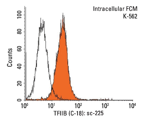
TFIIB (C-18): sc-225. Indirect, intracellular FCM analysis of fixed and permeabilized K-562 cells stained with TFIIB (C-18), followed by PE-conjugated goat anti-rabbit IgG: sc-3739. Black line histogram represents the isotype control, normal rabbit IgG: sc-3888.
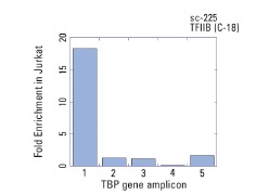
TFIIB (C-18): sc-225. ChIP analysis of TFIIB recruitment to genomic amplicons. Two (1,2) different human genomic TBP and three control amplicons (3-5) were analyzed by quantitative PCR (primer sequences available in on-line supplemental data). Data generated in collaboration with Drs. N. Trinklein and R. Myers, Stanford University (ENCODE Project).
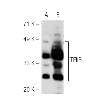
TFIIB (C-18): sc-225. Western blot analysis of TFIIB expression in non-transfected: sc-117752 (A) and mouse TFIIB transfected: sc-124000 (B) 293T whole cell lysates.
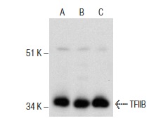
TFIIB (C-18): sc-225. Western blot analysis of TFIIB expression in HeLa (A), Hep G2 (B) and U-937 (C) nuclear extracts.
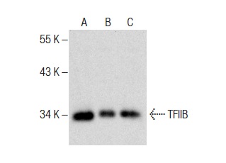
TFIIB (C-18): sc-225. Western blot analysis of TFIIB expression in Jurkat (A), K-562 (B) and A-431 (C) nuclear extracts.


