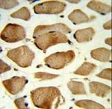
Formalin-fixed and paraffin-embedded human skeletal muscle reacted with CAV2 Antibody , which was peroxidase-conjugated to the secondary antibody, followed by DAB staining. This data demonstrates the use of this antibody for immunohistochemistry; clinical relevance has not been evaluated.
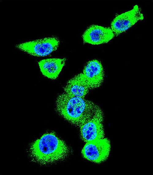
Confocal immunofluorescent of CAV2 Antibody with MDA-MB231 cell followed by Alexa Fluor 488-conjugated goat anti-rabbit lgG (green). DAPI was used to stain the cell nuclear (blue).

Western blot of lysate from HUVEC cell line,using CAV2 Antibody. Antibody was diluted at 1:1000 at each lane. A goat anti-rabbit IgG H&L (HRP) at 1:5000 dilution was used as the secondary antibody.Lysate at 35ug per lane.

CAV2 Antibody flow cytometry of MDA-MB231 cells (bottom histogram) compared to a negative control cell (top histogram). FITC-conjugated goat-anti-rabbit secondary antibodies were used for the analysis.