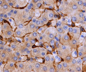
Caveolin-2 in Human Liver. Caveolin-2 was detected in paraffin-embedded sections of human liver using Goat Anti-Human/Mouse/Rat Caveolin-2 Antigen Affinity-purified Polyclonal Antibody at 10 ug/ml overnight at 4°C. Before incubation with the primary antibody tissue was subjected to heat-induced epitope retrieval using Antigen Retrieval Reagent-Basic. Tissue was stained using Anti-Goat HRP-DAB (brown) and counter-stained with hematoxylin (blue).
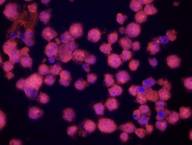
Caveolin-2 in Human K562 Cells. Caveolin2 was detected in immersion fixed K562 human chronic myelogenous leukemia cell line using 10 ug/ml Goat Anti-Human/ Mouse/Rat Caveolin2 Antigen Affinity-purified Polyclonal Antibody for 3 hours at room temperature. Cells were stained with the NorthernLights 557-conjugated Anti-Goat IgG Secondary Antibody (red ) and counterstained with DAPI (blue).
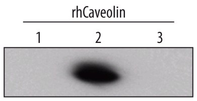
Detection of Human Caveolin-2 by Western Blot. Western blot shows recombinant human Caveolin-1, Caveolin-2 and Caveolin-3 (5 ng/lane). PVDF membrane was probed with 1 ug/ml Goat Anti-Human/Mouse/Rat Caveolin-2 Antigen Affinity-purified Polyclonal Antibody followed by HRP-conjugated Anti-Goat IgG Secondary Antibody. A specific band for Caveolin-2 was detected at approximately 22 kD (as indicated). This experiment was conducted under reducing conditions.
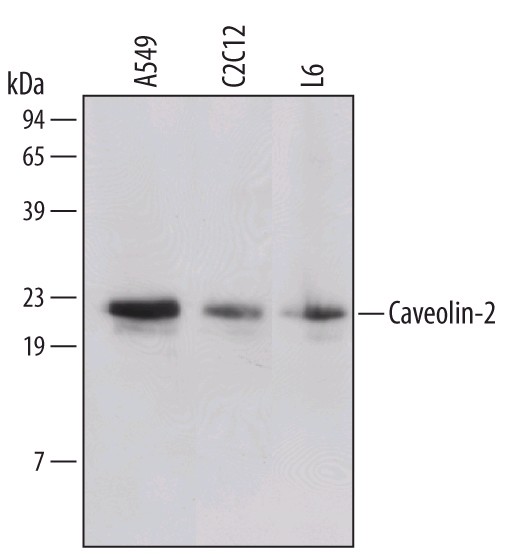
Detection of Human/Mouse/Rat Caveolin-2 by Western Blot. Western blot shows lysates of A549 human lung carcinoma cell line, C2C12 mouse myoblast cell line, and L6 rat myoblast cell line. PVDF membrane was probed with 1 ug /ml Goat Anti-Human/Mouse/Rat Caveolin-2 Antigen Affinity-purified Polyclonal Antibody followed by HRP-conjugated Anti-Goat IgG Secondary Antibody. A specific band for Caveolin-2 was detected at approximately 22 kD (as indicated). This experiment was conducted under reducing conditions.