Anti-CASP12 / Caspase 12 Antibody (aa165-193)
| Name | Anti-CASP12 / Caspase 12 Antibody (aa165-193) |
|---|---|
| Supplier | LifeSpan Bioscience |
| Catalog | LS-C168327 |
| Prices | $295.00 |
| Sizes | 400 µl |
| Host | Rabbit |
| Clonality | Polyclonal |
| Applications | IHC WB |
| Species Reactivities | Mouse, Human, Rat |
| Purity/Format | Protein A purified |
| Blocking Peptide | CASKIN2 Antibody Blocking Peptide |
| Description | Rabbit Polyclonal |
| Gene | CASP12 |
| Conjugate | Unconjugated |
| Supplier Page | Shop |
Product images
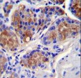 CASP12 Antibody immunohistochemistry of formalin-fixed and paraffin-embedded human stomach tissue followed by peroxidase-conjugated secondary antibody and DAB staining.
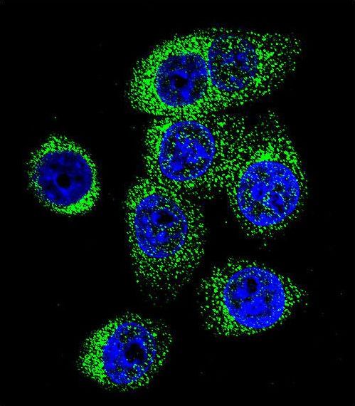 Confocal immunofluorescent of CASP12 Antibody with 293 cell followed by Alexa Fluor 488-conjugated goat anti-rabbit lgG (green). DAPI was used to stain the cell nuclear (blue).
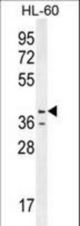 CASP12 Antibody western blot of HL-60 cell line lysates (35 ug/lane). The CASP12 antibody detected the CASP12 protein (arrow).
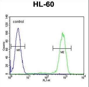 CASP12 Antibody flow cytometry of HL-60 cells (right histogram) compared to a negative control cell (left histogram). FITC-conjugated goat-anti-rabbit secondary antibodies were used for the analysis.