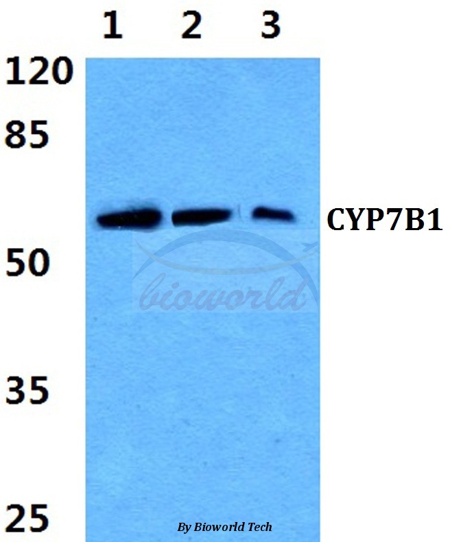
Western blot (WB) analysis of CYP7B1 (Q127) pAb at 1:500 dilution Lane1:DLD cell lysate Lane2:sp2/0 cell lysate Lane3:PC12 cell lysate
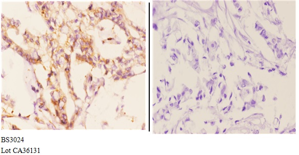
Immunohistochemistry (IHC) analyzes of CYP7B1 (Q127) pAb in paraffin-embedded human breast carcinoma tissue at 1:50.showing strong membranous and cytoplasmic staining. Negative control (the right)Using PBS instead of primary antibody, secondary antibody is Goat Anti-Rabbit IgG-biotin followed by avidin-peroxidase.
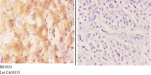
Immunohistochemistry (IHC) analyzes of CYP7B1 (Q127) pAb in paraffin-embedded human liver carcinoma tissue at 1:50.showing strong membranous and cytoplasmic staining. Negative control (the right)Using PBS instead of primary antibody, secondary antibody is
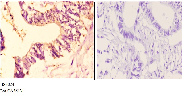
Immunohistochemistry (IHC) analyzes of CYP7B1 (Q127) pAb in paraffin-embedded human rectum carcinoma tissue at 1:50.showing strong membranous and cytoplasmic staining. Negative control (the right)Using PBS instead of primary antibody, secondary antibody i