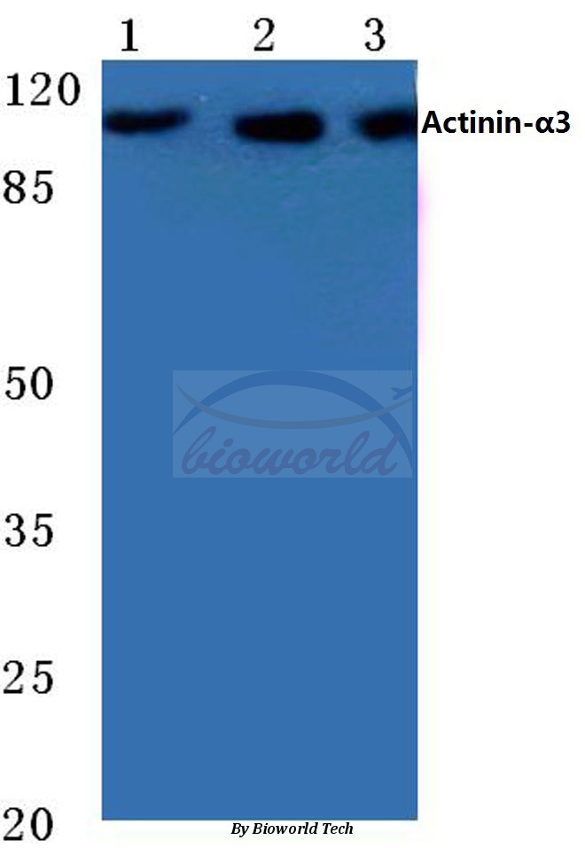
Western blot (WB) analysis of Actinin-α3 (E25) pAb at 1:500 dilution Lane1:Hela cell lysate Lane2:Mouse muscle tissue lysate Lane3:Rat muscel tissue lysate
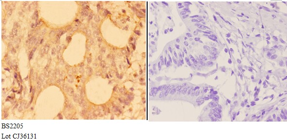
Immunohistochemistry (IHC) analyzes of Actinin-α3 (E25) pAb in paraffin-embedded human colon carcinoma tissue at 1:50,showing cytoplasmic and nucleus staining.Negative control (the right)Using PBS instead of primary antibody, secondary antibody is Goat Anti-Rabbit IgG-biotin followed by avidin-peroxidase.
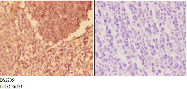
Immunohistochemistry (IHC) analyzes of Actinin-α3 (E25) pAb in paraffin-embedded human tonsil carcinoma tissue at 1:50,showing cytoplasmic and nucleus staining.Negative control (the right)Using PBS instead of primary antibody, secondary antibody is Goat A
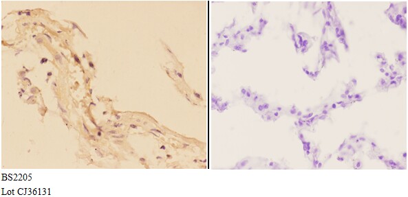
Immunohistochemistry (IHC) analyzes of Actinin-α3 (E25) pAb in paraffin-embedded human lung carcinoma tissue at 1:50,showing cytoplasmic and nucleus staining.Negative control (the right)Using PBS instead of primary antibody, secondary antibody is Goat Ant

Immunohistochemistry (IHC) analyzes of Actinin-α3 (E25) pAb in paraffin-embedded human kidney carcinoma tissue at 1:50,showing cytoplasmic and nucleus staining.Negative control (the right)Using PBS instead of primary antibody, secondary antibody is Goat A
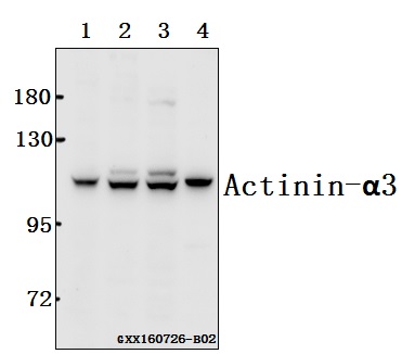
Western blot (WB) analysis of Actinin-α3 (E25) pAb at 1:500 dilution Lane1:MCF-7 whole cell lysate(40ug) Lane2:Hela whole cell lysate(40ug) Lane3:BV2 whole cell lysate(40ug) Lane4:C6 whole cell lysate(40ug)