KKIAMRE (L240) polyclonal antibody
| Name | KKIAMRE (L240) polyclonal antibody |
|---|---|
| Supplier | Bioworld Technology |
| Catalog | BS2113 |
| Prices | $158.00, $275.00 |
| Sizes | 50 µl, 100 µl |
| Host | Rabbit |
| Clonality | Polyclonal |
| Applications | WB IHC |
| Species Reactivities | Human, Mouse, Rat |
| Antigen | Synthetic peptide, corresponding to amino acids 210-260 of Human KKIAMRE. |
| Purity/Format | The antibody was affinity-purified from rabbit antiserum by affinity-chromatography using epitope-specific immunogen and the purity is > 95% (by SDS-PAGE). |
| Blocking Peptide | KKIAMRE (L240) Peptide |
| Description | Rabbit Polyclonal |
| Gene | CDKL2 |
| Supplier Page | Shop |
Product images
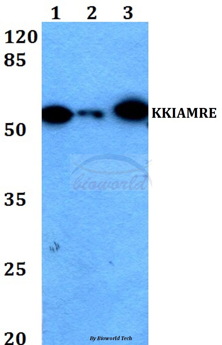 Western blot (WB) analysis of KKIAMRE (L240) pAb at 1:500 dilution Lane1:HEK293T whole cell lysate Lane2:Raw264.7 whole cell lysate Lane3:H9C2 whole cell lysate
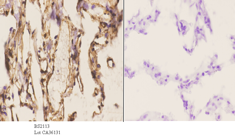 Immunohistochemistry (IHC) analyzes of KKIAMRE (L240) pAb in paraffin-embedded human lung carcinoma tissue at 1:50.showing cytoplasmic and nucleus staining. Negative control (the right)Using PBS instead of primary antibody, secondary antibody is Goat Anti-Rabbit IgG-biotin followed by avidin-peroxidase.
 Western blot (WB) analysis of KKIAMRE (L240) pAb at 1:500 dilution Lane1:HEK293T whole cell lysate(40ug) Lane2:786-O whole cell lysate(40ug)