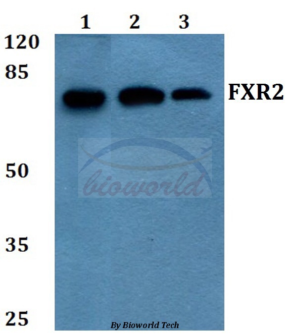
Western blot (WB) analysis of FXR2 (E576) pAb at 1:500 dilution Lane1:HEK293T whole cell lysate Lane2:NIH-3T3 whole cell lysate Lane3:H9C2 whole cell lysate
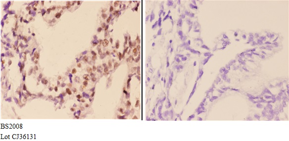
Immunohistochemistry (IHC) analyzes of FXR2 (E576) pAb in paraffin-embedded human colon carcinoma tissue at 1:50,showing cytoplasmic and nuclear staining.Negative control (the right)Using PBS instead of primary antibody, secondary antibody is Goat Anti-Rabbit IgG-biotin followed by avidin-peroxidase.
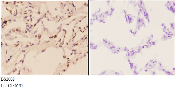
Immunohistochemistry (IHC) analyzes of FXR2 (E576) pAb in paraffin-embedded human lung carcinoma tissue at 1:50,showing cytoplasmic and nuclear staining.Negative control (the right) Using PBS instead of primary antibody, secondary antibody is Goat Anti-Ra
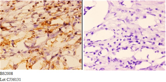
Immunohistochemistry (IHC) analyzes of FXR2 (E576) pAb in paraffin-embedded human kidney carcinoma tissue at 1:50,showing cytoplasmic and nuclear staining.Negative control (the right)Using PBS instead of primary antibody, secondary antibody is Goat Anti-R