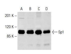
Sp1 (PEP 2)-G: sc-59-G. Western blot analysis of Sp1 expression in K-562 (A) and Jurkat (C) nuclear extracts and K-562 (B) and Jurkat (D) whole cell lysates.
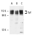
Sp1 (PEP 2): sc-59. Western blot analysis of Sp1 expression in HeLa (A), K-562 (B) and A-431 (C) whole cell lysates.
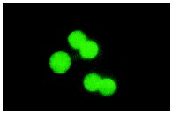
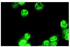
Sp1 (PEP 2): sc-59. Immunofluorescence staining of methanol-fixed KNRK cells showing nuclear localization.
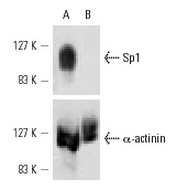
Sp1 siRNA (h): sc-29487. Western blot analysis of Sp1 expression in non-transfected control (A) and Sp1 siRNA transfected (B) A-431 cells. Blot probed with Sp1 (PEP 2): sc-59. α-actinin (H-2): sc-17829 used as specificity and loading control.
Sp1 siRNA (h): sc-29487. Immunofluorescence staining of methanol-fixed, control HeLa (A) and Sp1 siRNA silenced HeLa (B) cells showing diminished nuclear staining in the siRNA silenced cells. Cells probed with Sp1 (PEP 2): sc-59.
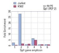
Sp1 (PEP-2): sc-59. ChIP analysis of Sp1 recruitment to genomic amplicons. Four (1-4) different human genomic Sp-1and two control amplicons (5,6) were analyzed by quantitative PCR (primer sequences available in on-line supplemental data). Data generated in collaboration with Drs. N. Trinklein and R. Myers, Stanford University (ENCODE Project).

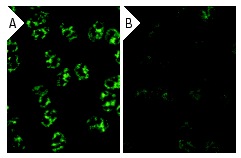
Sp1 (PEP 2): sc-59. Immunofluorescence staining of methanol-fixed NIH/3T3 cells showing nuclear localization using indirect FITC (A) staining and HeLa cells using direct Alexa Fluor 488 (B) staining.
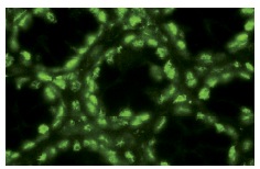
Sp1 (PEP 2): sc-59. Immunofluorescence staining of normal mouse intestine frozen section showing nuclear staining.
Sp1 (PEP 2)-G: sc-59-G. Immunofluorescence staining of methanol-fixed NIH/3T3 cells showing nuclear localization.

Sp1 (PEP 2)-G: sc-59-G. Immunoperoxidase staining of formalin-fixed, paraffin-embedded human colon carcinoma tissue showing nuclear localization.