RIPAb+ Upf1 - RIP Validated Antibody and Primer Set
| Name | RIPAb+ Upf1 - RIP Validated Antibody and Primer Set |
|---|---|
| Supplier | EMD Millipore |
| Catalog | 03-191 |
| Prices | $389.00 |
| Sizes | 10 assays 10 assays per set. recommended use: ~7 μl of antibody per rip . (dependent upon biological context) |
| Host | Rabbit |
| Clonality | Polyclonal |
| Applications | IP RIP-Chip WB |
| Species Reactivities | Dog, Bovine, Human, Mouse, Rat |
| Antigen | KLH-conjugated synthetic linear peptide corresponding to Upf1. |
| Purity/Format | Affinity Purfied |
| Description | Rabbit Polyclonal |
| Gene | UPF1 |
| Supplier Page | Shop |
Product images
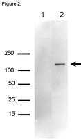 Immunoprecipitation from RIP lysate:
 Representative lot data.
RIP lysate from HeLa cells (~2 X 10E7 cell equivalents per IP) was subjected to immunoprecipitation using either 7 µL of a normal rabbit IgG or 7 µL of Anti-Upf1 antibody.
Ten percent of the precipitated proteins (lane 1: rabbit IgG, lane 2: Upf1) were resolved by electrophoresis, transferred to PVDF and probed with anti-Upf1 antibody (1:2000).
Proteins were visualized using One-Step™ IP-Western kit (GenScript Cat. # L00232)
Arrow indicates Upf1 (~125 kDa) (Figure 2).