Collagen Type IV Antibody Biotin Conjugated
| Name | Collagen Type IV Antibody Biotin Conjugated |
|---|---|
| Supplier | Rockland Immunochemicals Inc |
| Catalog | 600-406-106 |
| Prices | $319.00 |
| Sizes | 100 µg |
| Host | Rabbit |
| Clonality | Polyclonal |
| Applications | ELISA WB IHC |
| Species Reactivities | Human, Bovine |
| Antigen | Anti-Collagen Type IV was produced by repeated immunizations with Collagen Type IV from human and bovine placenta. |
| Purity/Format | Anti-Collagen Type IV has been prepared by immunoaffinity chromatography using immobilized antigens followed by extensive cross-adsorption against other collagens, human serum proteins and non-collagen extracellular matrix proteins to remove any unwanted specificities |
| Description | Rabbit Polyclonal |
| Gene | COL4A4 |
| Conjugate | Biotin |
| Supplier Page | Shop |
Product images
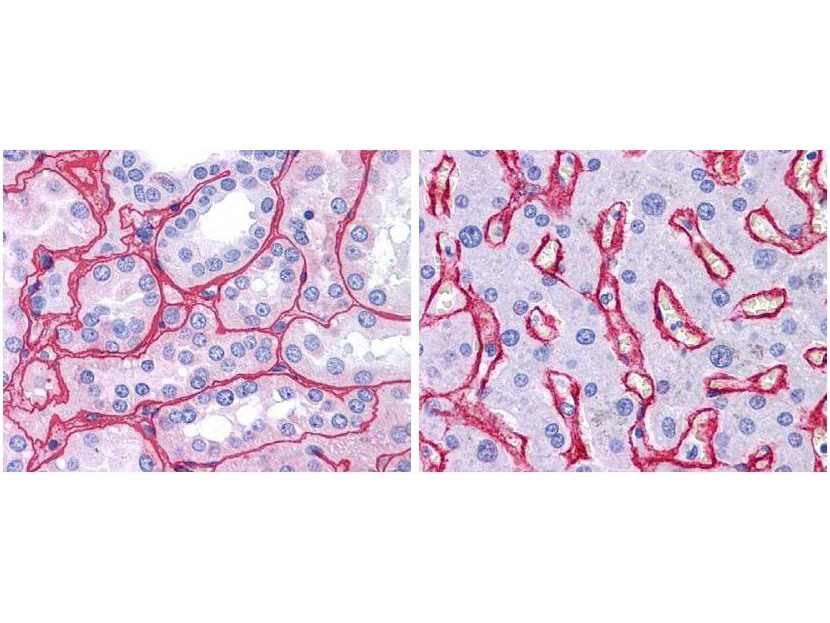 Immunohistochemistry of Rabbit Anti-Collagen IV Biotin Conjugated Antibody. Tissue: human kidney (Left) with strong red staining observed in glomeruli and liver (Right) with strong staining in sinusoids. Fixation: formalin fixed paraffin embedded. Antigen retrieval: steamed in 0.01 M sodium citrate buffer, pH 6.0 at 99-100°C - 20 minutes. Primary antibody: Collagen IV antibody at 10 µg/mL for 1 h at RT. Secondary antibody: Peroxidase rabbit secondary antibody at 1:10,000 for 45 min at RT. Localization: Collagen IV is extracellular. Staining: Collagen IV as precipitated red signal with hematoxylin purple nuclear counterstain.