Anti-BIRC2 / cIAP1 Antibody (aa2-18)
| Name | Anti-BIRC2 / cIAP1 Antibody (aa2-18) |
|---|---|
| Supplier | LifeSpan Bioscience |
| Catalog | LS-C148259 |
| Prices | $335.00 |
| Sizes | 50 µl |
| Host | Rabbit |
| Clonality | Polyclonal |
| Applications | WB ELISA |
| Species Reactivities | Human, Mouse, Rat |
| Antigen | BIRC2 / cIAP1 antibody was raised against a synthetic peptide corresponding to amino acids 2-18 (HKTASQRLFPGPSYQNI) of human cIAP1, GenBank no. gi|2497238. Percent identity by BLAST analysis: Human, Gorilla, Gibbon, Monkey (100%); Marmoset (88%). |
| Purity/Format | Antiserum |
| Description | Rabbit Polyclonal |
| Gene | BIRC2 |
| Conjugate | Unconjugated |
| Supplier Page | Shop |
Product images
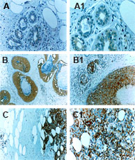 IHC of formalin-fixed, paraffin-embedded human breast tissue using LS-C148259 at 1:2000. A, normal breast tissue. B, ductal carcinoma in situ (DCIS). C, invasive breast carcinoma. A1, B1, and C1 are higher magnifications of A, B and C, respectively. Hematoxylin-Eos in counterstain. IncreasedcIAP1 expression is seen in the cancerous compared to normal breast tissue. Hematoxylin-Eos in counterstain.
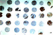 IHC of a formalin-fixed, paraffin-embedded human ovarian cancer tissue microarray using LS-C148259 at 1:2000. Differential expression of cIAP1 is seen between patient samples. Hematoxylin-Eosin counterstain.