Anti-BCL2L11 / BIM Antibody (aa134-160) IHC-plusâ¢
| Name | Anti-BCL2L11 / BIM Antibody (aa134-160) IHC-plus⢠|
|---|---|
| Supplier | LifeSpan Bioscience |
| Catalog | LS-B10712 |
| Prices | $395.00 |
| Sizes | 200 µl |
| Host | Rabbit |
| Clonality | Polyclonal |
| Applications | ICC/IF WB IP FC ELISA |
| Species Reactivities | Human, Monkey |
| Purity/Format | Protein A purified |
| Blocking Peptide | BCL2L1 / BCL-XL Antibody Blocking Peptide |
| Description | Rabbit Polyclonal |
| Gene | BCL2L11 |
| Conjugate | Unconjugated |
| Supplier Page | Shop |
Product images
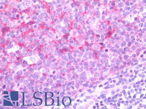 Anti-BCL2L11 / BIM antibody IHC staining of human tonsil. Immunohistochemistry of formalin-fixed, paraffin-embedded tissue after heat-induced antigen retrieval. Antibody LS-B10712 dilution 1:50.
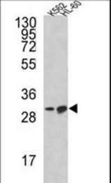 Western blot of BCL2L11 Antibody in K562, HL-60 cell line lysates (35 ug/lane). BCL2L11 (arrow) was detected using the purified antibody.
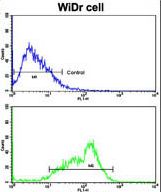 Flow cytometric of WiDr cells using BCL2L11 Antibody (bottom histogram) compared to a negative control cell (top histogram). FITC-conjugated goat-anti-rabbit secondary antibodies were used for the analysis.