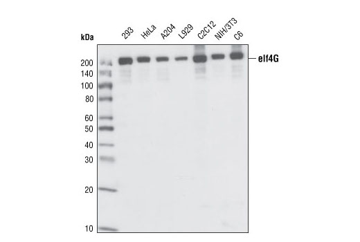
Western blot analysis of extracts from various cell lines, using eIF4G Antibody.

Immunohistochemical analysis of paraffin-embedded human colon carcinoma, showing cytoplasmic localization, using eIF4G Antibody.
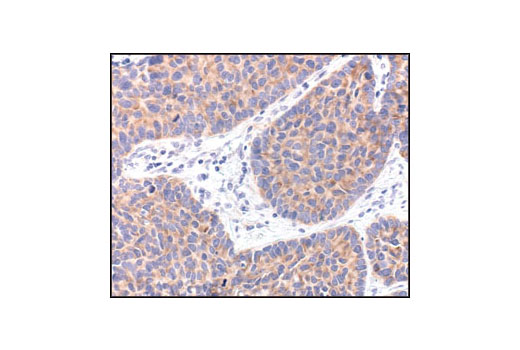
Immunohistochemical analysis of paraffin-embedded human lung carcinoma, showing cytoplasmic localization, using eIF4G Antibody.

Immunohistochemical analysis of paraffin-embedded human breast carcinoma, using eIF4G Antibody in the presence of control peptide (left) or eIF4G blocking peptide #1003 (right).

Confocal immunofluorescent analysis of HeLa cells, using eIF4G Antibody (green, left) compared to an isotype control (right). Actin filaments have been labeled with Alexa Fluor® 555 phalloidin (red). Blue pseudocolor = DRAQ5 ® #4084 (fluorescent DNA dye).

Flow cytometric analysis of HeLa cells, using eIF4G Antibody (blue) compared to a nonspecific negative control antibody (red).