Anti-AUTL1 / ATG4C Antibody (aa40-70)
| Name | Anti-AUTL1 / ATG4C Antibody (aa40-70) |
|---|---|
| Supplier | LifeSpan Bioscience |
| Catalog | LS-C156600 |
| Prices | $295.00 |
| Sizes | 400 µl |
| Host | Rabbit |
| Clonality | Polyclonal |
| Applications | IHC-P WB |
| Species Reactivities | Human, Mouse |
| Antigen | AUTL1 / ATG4C antibody was raised against kLH-conjugated synthetic peptide from N-terminal of human APG4C. |
| Purity/Format | Immunoaffinity purified |
| Blocking Peptide | AURKC / Aurora C Antibody Blocking Peptide |
| Description | Rabbit Polyclonal |
| Gene | ATG4C |
| Supplier Page | Shop |
Product images
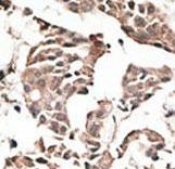 Formalin-fixed and paraffin-embedded human cancer tissue reacted with the primary antibody, which was peroxidase-conjugated to the secondary antibody, followed by AEC staining. This data demonstrates the use of this antibody for immunohistochemistry; clinical relevance has not been evaluated. BC = breast carcinoma; HC = hepatocarcinoma.
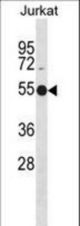 APG4C Antibody (Y48) western blot of Jurkat cell line lysates (35 ug/lane). The APG4C antibody detected the APG4C protein (arrow).
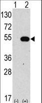 Western blot of anti-hAPG4C-Y48 antibody in 293 cell line lysates transiently transfected with the ATG4C gene (2 ug/lane). hAPG4C-Y48(arrow) was detected using the purified antibody.
 The anti-APG4C antibody is used in Western blot to detect APG4C in mouse liver tissue lysate