Anti-ASMT / HIOMT Antibody (aa219-249)
| Name | Anti-ASMT / HIOMT Antibody (aa219-249) |
|---|---|
| Supplier | LifeSpan Bioscience |
| Catalog | LS-C156543 |
| Prices | $295.00 |
| Sizes | 400 µl |
| Host | Rabbit |
| Clonality | Polyclonal |
| Applications | IHC-P WB FC |
| Species Reactivities | Human |
| Antigen | ASMT / HIOMT antibody was raised against kLH-conjugated synthetic peptide from internal region of human ASMT. |
| Purity/Format | Immunoaffinity purified |
| Blocking Peptide | ASH2L / ASH2 Antibody Blocking Peptide |
| Description | Rabbit Polyclonal |
| Gene | ASMT |
| Supplier Page | Shop |
Product images
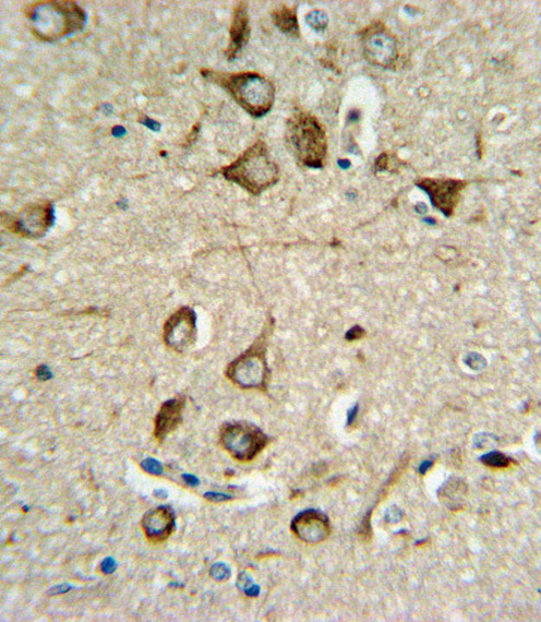 ASMT Antibody IHC of formalin-fixed and paraffin-embedded brain tissue followed by peroxidase-conjugated secondary antibody and DAB staining.
 Western blot of ASMT Antibody in 293 cell line lysates (35 ug/lane). ASMT (arrow) was detected using the purified antibody.
 Western blot of lysate from Y79 cell line, using ASMT Antibody. Antibody was diluted at 1:1000. A goat anti-rabbit IgG H&L (HRP) at 1:10000 dilution was used as the secondary antibody. Lysate at 20ug.
 ASMT Antibody flow cytometry of 293 cells (right histogram) compared to a negative control cell (left histogram). FITC-conjugated goat-anti-rabbit secondary antibodies were used for the analysis.