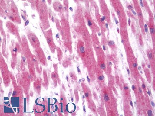
Anti-ALOX12/12 Lipoxygenase/12-LOX antibody IHC staining of human heart. Immunohistochemistry of formalin-fixed, paraffin-embedded tissue after heat-induced antigen retrieval. Antibody LS-B10714 dilution 1:50.
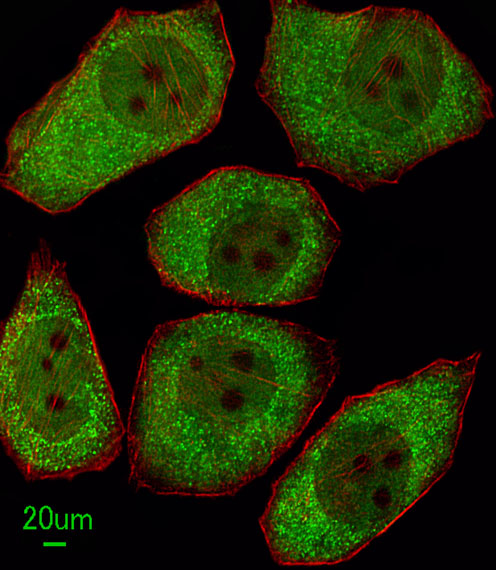
Immunofluorescent of A549 cells, using ALOX12 Antibody. Antibody was diluted at 1:100 dilution. Alexa Fluor 488-conjugated goat anti-rabbit lgG at 1:400 dilution was used as the secondary antibody (green). Cytoplasmic actin was counterstained with Dylight Fluor 554 (red) conjugated Phalloidin (red).
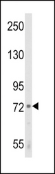
Western blot of ALOX12 Antibody in K562 cell line lysates (35 ug/lane). ALOX12 (arrow) was detected using the purified antibody.
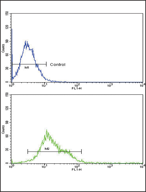
ALOX12 Antibody flow cytometry of k562 cells (bottom histogram) compared to a negative control cell (top histogram). FITC-conjugated goat-anti-rabbit secondary antibodies were used for the analysis.