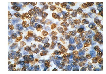
PML (PG-M3): sc-966. Immunoperoxidase staining of formalin-fixed, paraffin-embedded normal human lymph node showing nuclear staining.
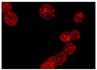
PML (PG-M3): sc-966. Immunofluorescence staining of methanol-fixed K-562 cells showing nuclear staining.
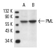
PML (PG-M3): sc-966. Western blot analysis of PML expression in K-562 (A) and COLO 320DM (B) whole cell lysates.
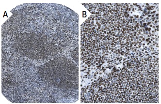
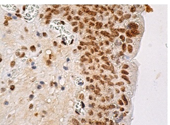
PML (PG-M3): sc-966. Immunoperoxidase staining of formalin fixed, paraffin-embedded human lymph node tissue showing nuclear staining of lymphoid cells (low and high magnification). Kindly provided by The Swedish Human Protein Atlas (HPA) program.
PML (PG-M3): sc-966. Immunoperoxidase staining of formalin fixed, paraffin-embedded human urinary bladder tissue showing speckled staining of nucleus in urothelial cells.