
p300 (N-15): sc-584. Western blot analysis of p300 expression in A-431 nuclear extract.
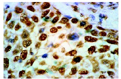
p300 (N-15): sc-584. Immunoperoxidase staining of formalin-fixed, paraffin-embedded human breast carcinoma tissue showing nuclear localization.
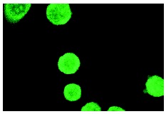
p300 (N-15): sc-584. Immunofluorescence staining of methanol-fixed SK-BR-3 cells showing nuclear localization.
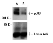
p300 siRNA (h): sc-29431. Western blot analysis of p300 expression in non-transfected control (A) and p300 siRNA transfected (B) A-431 cells. Blot probed with p300 (N-15): sc-584. Lamin A/C (636): sc-7292 used as specificity and loading control.
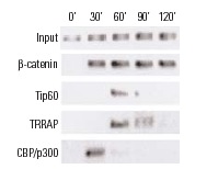
ChIP analysis of coactivator recruitment on Cyclin D2 promoter in C2C12 cells treated with LiCl and serum. Antibodies tested include β-catenin (H-102): sc-7199, β-catenin (C-18): sc-1496, β-catenin (E-5): sc-7963, Tip60 (N-17): sc-5725, TRRAP (T-17): sc-5405, TRRAP (Y-18): sc-12375, TRRAP (F-20): sc-12376, TRRAP (H-300): sc-11411, CBP (A-22): sc-369, CBP (C-20): sc-583, CBP (451): sc-1211, CPB (C-1): sc-7300, p300 (H-272): sc-8981, p300 (N-15): sc-584 and p300 (C-20): sc-585. Data kindly provided by M.G. Rosenfeld and reproduced with permission from Kioussi et al., Cell 2002, 111: 673-685.

ChIP analysis of cytokine gene promoter occupancy in TNFα-treated mouse embryonic fibroblasts. IP's permorned without (A) and with (B) primary antibody. Antibodies tested include NFκB p65 (A): sc-109, RelB (C-19): sc-226 and p300 (N-15): sc-584. Data kindly provided by A. Hoffmann.
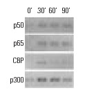
ChIP analysis of cofactor occupancy dynamics on the IL-8 promoter in 293 cells in response to IL-1β treatment. Antibodies tested include NFκB p50 (C-19): sc-1190, NFκB p50 (E-10): sc-8414, NFκB p50 (H-119): sc-7178, NFκB p65 (C-20): sc-372, NFκB p65 (A): sc-109, NFκB p65 (H-286): sc-7151, CBP (A-22): sc-369, CBP (C-1): sc-7300, CBP (C-20): sc-583, CBP (451): sc-1211, p300 (C-20): sc-sc-585, p300 (N-15): sc-584, p300 (H-272): sc-8981. Data kindly provided by M.G. Rosenfeld and reproduced with permission from Baek et al., Cell 2002, 110: 55-67.
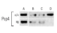
ChIP analysis of in vivo binding of RORα and its recruitment of coactivators to RORα-responsive promoters in freshly dissected cerebella derived from wild type (+/+) and staggerer (Sg) mice. Control Input (A). Antibodies used included TRβ1 (J51): sc-737 and TRβ1 (J52): sc-738 (B), p300 (C-20): sc-585, p300 (H-272): sc-8981 and p300 (N-15): sc-584 (C), β-catenin (H-102): sc-7199, β-catenin (C-18): sc-1496 and β-catenin (E-5): sc-7963 (D). Data kindly provided by M.G. Rosenfeld and reproduced wtih permission from Gold et al., Neuron 2003, 40: 1119-1131.
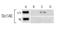
ChIP analysis of in vivo binding of RORα and its recruitment of coactivators to RORα-responsive promoters in freshly dissected cerebella derived from wild type (+/+) and staggerer (Sg) mice. Control Input (A). Antibodies used included TRβ1 (J51): sc-737 and TRβ1 (J52): sc-738 (B), p300 (C-20): sc-585, p300 (H-272): sc-8981 and p300 (N-15): sc-584 (C), β-catenin (H-102): sc-7199, β-catenin (C-18): sc-1496 and β-catenin (E-5): sc-7963 (D). Data kindly provided by M.G. Rosenfeld and reproduced wtih permission from Gold et al., Neuron 2003, 40: 1119-1131.

ChIP analysis of in vivo binding of RORα and its recruitment of coactivators to RORα-responsive promoters in freshly dissected cerebella derived from wild type (+/+) and staggerer (Sg) mice. Control Input (A). Antibodies used included TRβ1 (J51): sc-737 and TRβ1 (J52): sc-738 (B), p300 (C-20): sc-585, p300 (H-272): sc-8981 and p300 (N-15): sc-584 (C), β-catenin (H-102): sc-7199, β-catenin (C-18): sc-1496 and β-catenin (E-5): sc-7963 (D). Data kindly provided by M.G. Rosenfeld and reproduced wtih permission from Gold et al., Neuron 2003, 40: 1119-1131.
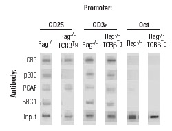
ChIP analysis of recruitment of CBP, p300, PCA and BRG1 to the IL-2α (CD25) promoter in vivo. Antibodies tested include CBP (A-22): sc-369, p300 (N-15): sc-584, PCAF (H-369): sc-8999, and Brg-1 (H-88): sc-10768. CD3ε and Oct-2 promoter regions were employed as positive and negative controls, respectively. DNA was isolated from Rag
-/- thymocytes or Rag
-/- thymocytes expressing TCRβ. Data kindly provided by J. Imbert and reproduced from Yeh, J-H., et al., Nucleic Acids Research, 2002, 30: 1944-1951, with permission from Oxford University Press.
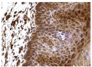
p300 (N-15): sc-584. Immunoperoxidase staining of formalin fixed, paraffin-embedded human esophagus tissue showing nuclear and cytoplasmic staining of squamous epithelial cells.


