Anti-ADI1 / ARD Antibody (clone 3H6)
| Name | Anti-ADI1 / ARD Antibody (clone 3H6) |
|---|---|
| Supplier | LifeSpan Bioscience |
| Catalog | LS-C115667 |
| Prices | $325.00 |
| Sizes | 100 µl |
| Host | Mouse |
| Clonality | Monoclonal |
| Isotype | IgG1 |
| Clone | 3H6 |
| Applications | ICC/IF WB FC |
| Species Reactivities | Human |
| Antigen | ADI1 / ARD antibody was raised against full length human recombinant protein of human ADI1 (NP_060739) produced in HEK293T cell. |
| Purity/Format | Protein A/G purified |
| Description | Mouse Monoclonal |
| Gene | ADI1 |
| Conjugate | Unconjugated |
| Supplier Page | Shop |
Product images
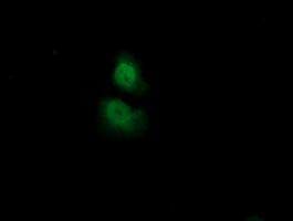 Anti-ADI1 mouse monoclonal antibody immunofluorescent staining of COS7 cells transiently transfected by pCMV6-ENTRY ADI1.
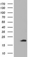 HEK293T cells were transfected with the pCMV6-ENTRY control (Left lane) or pCMV6-ENTRY ADI1 (Right lane) cDNA for 48 hrs and lysed. Equivalent amounts of cell lysates (5 ug per lane) were separated by SDS-PAGE and immunoblotted with anti-ADI1.
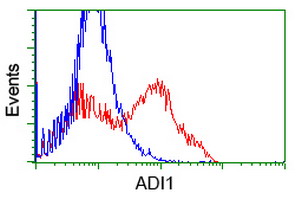 HEK293T cells transfected with either overexpress plasmid (Red) or empty vector control plasmid (Blue) were immunostained by anti-ADI1 antibody, and then analyzed by flow cytometry.