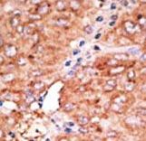
Formalin-fixed and paraffin-embedded human cancer tissue reacted with the primary antibody, which was peroxidase-conjugated to the secondary antibody, followed by AEC staining. This data demonstrates the use of this antibody for immunohistochemistry; clinical relevance has not been evaluated. BC = breast carcinoma; HC = hepatocarcinoma.
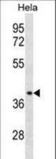
CCBP2 Antibody (A313) western blot of HeLa cell line lysates (35 ug/lane). The CCBP2 antibody detected the CCBP2 protein (arrow).
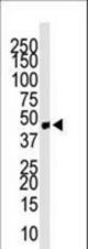
Western blot of anti-CCBP2 antibody in mouse small intestine tissue lysate. CCBP2 (Arrow) was detected using purified antibody. Secondary HRP-anti-rabbit was used for signal visualization with chemiluminescence.