IHC of paraffin-embedded Carcinoma of Human thyroid tissue using anti-ACAA2 mouse monoclonal antibody.

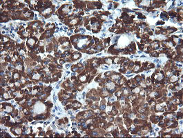
IHC of paraffin-embedded Human pancreas tissue using anti-ACAA2 mouse monoclonal antibody.

IHC of paraffin-embedded Human Kidney tissue using anti-ACAA2 mouse monoclonal antibody.

IHC of paraffin-embedded Human liver tissue using anti-ACAA2 mouse monoclonal antibody.

IHC of paraffin-embedded Human endometrium tissue using anti-ACAA2 mouse monoclonal antibody.

Anti-ACAA2 mouse monoclonal antibody immunofluorescent staining of COS7 cells transiently transfected by pCMV6-ENTRY ACAA2.

HEK293T cells were transfected with the pCMV6-ENTRY control (Left lane) or pCMV6-ENTRY ACAA2 (Right lane) cDNA for 48 hrs and lysed. Equivalent amounts of cell lysates (5 ug per lane) were separated by SDS-PAGE and immunoblotted with anti-ACAA2.
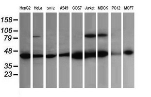
Western blot of extracts (35 ug) from 9 different cell lines by using anti-ACAA2 monoclonal antibody (HepG2: human; HeLa: human; SVT2: mouse; A549: human; COS7: monkey; Jurkat: human; MDCK: canine; PC12: rat; MCF7: human).