Anti-ABL1 / c-ABL Antibody (aa741-769)
| Name | Anti-ABL1 / c-ABL Antibody (aa741-769) |
|---|---|
| Supplier | LifeSpan Bioscience |
| Catalog | LS-C99162 |
| Prices | $295.00 |
| Sizes | 400 µl |
| Host | Rabbit |
| Clonality | Polyclonal |
| Applications | IHC-P ICC/IF WB IP |
| Species Reactivities | Human, Mouse |
| Purity/Format | Ammonium sulfate precipitation |
| Blocking Peptide | ABL Antibody Blocking Peptide |
| Description | Rabbit Polyclonal |
| Gene | ABL1 |
| Conjugate | Unconjugated |
| Supplier Page | Shop |
Product images
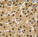 Formalin-fixed and paraffin-embedded human hepatocarcinoma tissue reacted with ABL1 antibody , which was peroxidase-conjugated to the secondary antibody, followed by DAB staining. This data demonstrates the use of this antibody for immunohistochemistry; clinical relevance has not been evaluated.
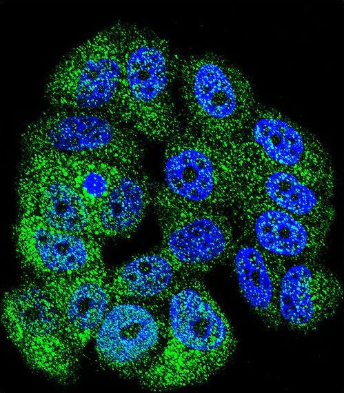 Confocal immunofluorescent of ABL1 Antibody with MCF-7 cell followed by Alexa Fluor 488-conjugated goat anti-rabbit lgG (green). DAPI was used to stain the cell nuclear (blue).
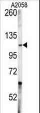 Western blot of anti-ABL1 Antibody in A2058 cell line lysates (35 ug/lane). ABL1 (arrow) was detected using the purified antibody.