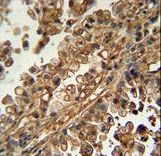
ABCG1 Antibody immunohistochemistry of formalin-fixed and paraffin-embedded human lung carcinoma followed by peroxidase-conjugated secondary antibody and DAB staining.
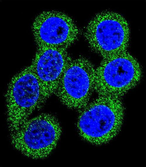
Confocal immunofluorescent of ABCG1 Antibody with 293 cell followed by Alexa Fluor 488-conjugated goat anti-rabbit lgG (green). DAPI was used to stain the cell nuclear (blue).
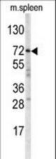
Western blot of ABCG1 antibody in mouse spleen tissue lysates (35 ug/lane). ABCG1 (arrow) was detected using the purified antibody.
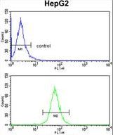
ABCG1 Antibody flow cytometry of HepG2 cells (bottom histogram) compared to a negative control cell (top histogram). FITC-conjugated goat-anti-rabbit secondary antibodies were used for the analysis.